 Movie
Movie Controller

[English] 日本語
 Yorodumi
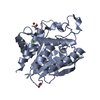
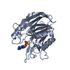
- PDB-5fyr: Calcium-dependent phosphoinositol-specific phospholipase C from a... -
+ Open data

- Basic information
| Entry | Database: PDB / ID: 5fyr | ||||||
|---|---|---|---|---|---|---|---|
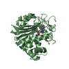
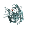
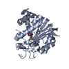
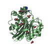
| Title | Calcium-dependent phosphoinositol-specific phospholipase C from a Gram-negative bacterium, Pseudomonas sp, apo form, myoinositol complex | ||||||
 Components Components | PHOSPHOINOSITOL-SPECIFIC PHOSPHOLIPASE C | ||||||
 Keywords Keywords | HYDROLASE / PI-PLC / BACTERIAL / PSEUDOMONAS / GRAM-NEGATIVE / CALCIUM-DEPENDENT / MYOINOSITOL COMPLEX | ||||||
| Function / homology | Phosphoinositide phospholipase C, Ca2+-dependent / Phosphoinositide phospholipase C, Ca2+-dependent / PLC-like phosphodiesterase, TIM beta/alpha-barrel domain superfamily / phosphoric diester hydrolase activity / lipid metabolic process / metal ion binding / 1,2,3,4,5,6-HEXAHYDROXY-CYCLOHEXANE / PHOSPHATE ION / Phosphoinositol-specific phospholipase c Function and homology information Function and homology information | ||||||
| Biological species |  PSEUDOMONAS SP. (bacteria) PSEUDOMONAS SP. (bacteria) | ||||||
| Method |  X-RAY DIFFRACTION / SYNCHROTRON / MOLECULAR REPLACEMENT / Resolution: 1.45 Å X-RAY DIFFRACTION / SYNCHROTRON / MOLECULAR REPLACEMENT / Resolution: 1.45 Å | ||||||
 Authors Authors | Moroz, O.V. / Blagova, E. / Lebedev, A.A. / Norgaard, A. / Segura, D.R. / Blicher, T.H. / Wilson, K.S. | ||||||
 Citation Citation | Journal: Acta Crystallogr D Struct Biol / Year: 2017 Title: The structure of a calcium-dependent phosphoinositide-specific phospholipase C from Pseudomonas sp. 62186, the first from a Gram-negative bacterium. Authors: Moroz, O.V. / Blagova, E. / Lebedev, A.A. / Nrgaard, A. / Segura, D.R. / Blicher, T.H. / Brask, J. / Wilson, K.S. | ||||||
| History |
|
- Structure visualization
| Structure viewer | Molecule: MolmilJmol/JSmol |
|---|
- Downloads & links
-Download
| PDBx/mmCIF format | 5fyr.cif.gz | 502.8 KB | Display | PDBx/mmCIF format |
|---|---|---|---|---|
| PDB format | pdb5fyr.ent.gz | 414.4 KB | Display | PDB format |
| PDBx/mmJSON format | 5fyr.json.gz | Tree view | PDBx/mmJSON format | |
| Others |  Other downloads Other downloads |
-Validation report
| Arichive directory | https://data.pdbj.org/pub/pdb/validation_reports/fy/5fyrftp://data.pdbj.org/pub/pdb/validation_reports/fy/5fyr | HTTPS FTP |
|---|
-Related structure data
| Related structure data |  5fyoC  5fypSC C: citing same article ( S: Starting model for refinement |
|---|---|
| Similar structure data |


-Links
 PDBj
PDBj- Assembly
| Deposited unit | 
| ||||||||
|---|---|---|---|---|---|---|---|---|---|
| 1 | 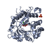
| ||||||||
| 2 | 
| ||||||||
| 3 | 
| ||||||||
| 4 | 
| ||||||||
| Unit cell |
|
-Components
| #1: Protein | Mass: 32775.258 Da / Num. of mol.: 4 Source method: isolated from a genetically manipulated source Source: (gene. exp.) PSEUDOMONAS SP. (bacteria) / Production host: #2: Chemical | ChemComp-INS /   Mass: 180.156 Da / Num. of mol.: 4 / Source method: obtained synthetically / Formula: C6H12O6 Mass: 180.156 Da / Num. of mol.: 4 / Source method: obtained synthetically / Formula: C6H12O6#3: Chemical | ChemComp-CA /   Mass: 40.078 Da / Num. of mol.: 4 / Source method: obtained synthetically / Formula: Ca Mass: 40.078 Da / Num. of mol.: 4 / Source method: obtained synthetically / Formula: Ca#4: Chemical |   Mass: 94.971 Da / Num. of mol.: 3 / Source method: obtained synthetically / Formula: PO4 Mass: 94.971 Da / Num. of mol.: 3 / Source method: obtained synthetically / Formula: PO4#5: Water | ChemComp-HOH / |  Mass: 18.015 Da / Num. of mol.: 1072 / Source method: isolated from a natural source / Formula: H2O Mass: 18.015 Da / Num. of mol.: 1072 / Source method: isolated from a natural source / Formula: H2OHas protein modification | Y | |
|---|
-Experimental details
-Experiment
| Experiment | Method: X-RAY DIFFRACTION / Number of used crystals: 1 |
|---|
- Sample preparation
| Crystal | Density Matthews: 1.99 Å3/Da / Density % sol: 38.4 % / Description: NONE |
|---|---|
| Crystal grow | Method: vapor diffusion, hanging drop / pH: 10 Details: PEG3350 24-27%, CAPS/BICINE MIX PH 9.0-10 GRID, BEST CRYSTALS CLOSER TO PH 10, SEEDING FROM PACT H4 (0.2M KSCN, 20% PEG3350, BIS TRIS PROPANE PH 8.5), ORYX ROBOT, 24 WELL PLATE, HANGING DROPS |
-Data collection
| Diffraction | Mean temperature: 100 K |
|---|---|
| Diffraction source | Source: SYNCHROTRON / Site: Diamond  / Beamline: I03 / Wavelength: 0.98 / Beamline: I03 / Wavelength: 0.98 |
| Detector | Type: DECTRIS PILATUS3 6M / Detector: PIXEL / Date: May 7, 2015 |
| Radiation | Protocol: SINGLE WAVELENGTH / Monochromatic (M) / Laue (L): M / Scattering type: x-ray |
| Radiation wavelength | Wavelength: 0.98 Å / Relative weight: 1 |
| Reflection | Resolution: 1.45→48.93 Å / Num. obs: 184175 / % possible obs: 99.7 % / Observed criterion σ(I): 2 / Redundancy: 7.2 % / Rmerge(I) obs: 0.07 / Net I/σ(I): 14.5 |
| Reflection shell | Resolution: 1.45→1.47 Å / Redundancy: 5.7 % / Rmerge(I) obs: 0.6 / Mean I/σ(I) obs: 2.5 / % possible all: 95.2 |
- Processing
| Software |
| ||||||||||||||||||||||||||||||||||||||||||||||||||||||||||||||||||||||||||||||||||||||||||||||||||||||||||||||||||||||||||||||||||||||||||||||||||||||||||||||||||||||||||||||||||||||
|---|---|---|---|---|---|---|---|---|---|---|---|---|---|---|---|---|---|---|---|---|---|---|---|---|---|---|---|---|---|---|---|---|---|---|---|---|---|---|---|---|---|---|---|---|---|---|---|---|---|---|---|---|---|---|---|---|---|---|---|---|---|---|---|---|---|---|---|---|---|---|---|---|---|---|---|---|---|---|---|---|---|---|---|---|---|---|---|---|---|---|---|---|---|---|---|---|---|---|---|---|---|---|---|---|---|---|---|---|---|---|---|---|---|---|---|---|---|---|---|---|---|---|---|---|---|---|---|---|---|---|---|---|---|---|---|---|---|---|---|---|---|---|---|---|---|---|---|---|---|---|---|---|---|---|---|---|---|---|---|---|---|---|---|---|---|---|---|---|---|---|---|---|---|---|---|---|---|---|---|---|---|---|---|
| Refinement | Method to determine structure: MOLECULAR REPLACEMENT Starting model: PDB ENTRY 5FYP Resolution: 1.45→48.93 Å / Cor.coef. Fo:Fc: 0.967 / Cor.coef. Fo:Fc free: 0.953 / SU B: 2.343 / SU ML: 0.041 / Cross valid method: THROUGHOUT / ESU R: 0.076 / ESU R Free: 0.069 / Stereochemistry target values: MAXIMUM LIKELIHOOD Details: HYDROGENS HAVE BEEN ADDED IN THE RIDING POSITIONS. U VALUES REFINED INDIVIDUALLY
| ||||||||||||||||||||||||||||||||||||||||||||||||||||||||||||||||||||||||||||||||||||||||||||||||||||||||||||||||||||||||||||||||||||||||||||||||||||||||||||||||||||||||||||||||||||||
| Solvent computation | Ion probe radii: 0.8 Å / Shrinkage radii: 0.8 Å / VDW probe radii: 1.2 Å / Solvent model: MASK | ||||||||||||||||||||||||||||||||||||||||||||||||||||||||||||||||||||||||||||||||||||||||||||||||||||||||||||||||||||||||||||||||||||||||||||||||||||||||||||||||||||||||||||||||||||||
| Displacement parameters | Biso mean: 16.818 Å2
| ||||||||||||||||||||||||||||||||||||||||||||||||||||||||||||||||||||||||||||||||||||||||||||||||||||||||||||||||||||||||||||||||||||||||||||||||||||||||||||||||||||||||||||||||||||||
| Refinement step | Cycle: LAST / Resolution: 1.45→48.93 Å
| ||||||||||||||||||||||||||||||||||||||||||||||||||||||||||||||||||||||||||||||||||||||||||||||||||||||||||||||||||||||||||||||||||||||||||||||||||||||||||||||||||||||||||||||||||||||
| Refine LS restraints |
|