 Movie
Movie Controller


+ Open data

- Basic information
| Entry | Database: PDB / ID: 7oml | ||||||
|---|---|---|---|---|---|---|---|
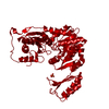
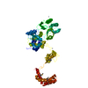
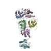
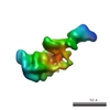
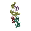
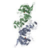
| Title | Bacillus subtilis phosphoglucomutase GlmM (metal bound) | ||||||
 Components Components | Phosphoglucosamine mutase | ||||||
 Keywords Keywords | ISOMERASE / Phosphoglucosamine Mutase / glucosamine-6-phosphate / glucosamine-1-phosphate / c-di-AMP / diadenylate cyclase / metal-bound protein. | ||||||
| Function / homology |  Function and homology information Function and homology informationphosphoglucosamine mutase / phosphoglucosamine mutase activity / phosphomannomutase activity / UDP-N-acetylglucosamine biosynthetic process / peptidoglycan biosynthetic process / carbohydrate metabolic process / magnesium ion binding / cytosol Similarity search - Function | ||||||
| Biological species |  | ||||||
| Method |  X-RAY DIFFRACTION / SYNCHROTRON / MOLECULAR REPLACEMENT / Resolution: 2.9 Å X-RAY DIFFRACTION / SYNCHROTRON / MOLECULAR REPLACEMENT / Resolution: 2.9 Å | ||||||
 Authors Authors | Pathania, M. / Grundling, A.G. / Freemont, P. | ||||||
| Funding support |  United Kingdom, 1items United Kingdom, 1items
| ||||||
 Citation Citation | Journal: J.Biol.Chem. / Year: 2021 Title: Structural basis for the inhibition of the Bacillus subtilis c-di-AMP cyclase CdaA by the phosphoglucomutase GlmM. Authors: Pathania, M. / Tosi, T. / Millership, C. / Hoshiga, F. / Morgan, R.M.L. / Freemont, P.S. / Grundling, A. | ||||||
| History |
|
- Structure visualization
| Structure viewer | Molecule: MolmilJmol/JSmol |
|---|
- Downloads & links
-Download
| PDBx/mmCIF format | 7oml.cif.gz | 97.1 KB | Display | PDBx/mmCIF format |
|---|---|---|---|---|
| PDB format | pdb7oml.ent.gz | 72.7 KB | Display | PDB format |
| PDBx/mmJSON format | 7oml.json.gz | Tree view | PDBx/mmJSON format | |
| Others |  Other downloads Other downloads |
-Validation report
| Arichive directory | https://data.pdbj.org/pub/pdb/validation_reports/om/7omlftp://data.pdbj.org/pub/pdb/validation_reports/om/7oml | HTTPS FTP |
|---|
-Related structure data
| Related structure data |  7ojrC 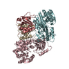 7ojsC 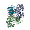 7olhC  3pdkS S: Starting model for refinement C: citing same article ( |
|---|---|
| Similar structure data | |
| Other databases |
|


-Links
 PDBj
PDBj- Assembly
| Deposited unit | 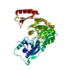
| ||||||||
|---|---|---|---|---|---|---|---|---|---|
| 1 | 
| ||||||||
| Unit cell |
|
-Components
| #1: Protein | Mass: 50314.867 Da / Num. of mol.: 1 Source method: isolated from a genetically manipulated source Source: (gene. exp.) Gene: glmM, ybbT, BSU01770 / Production host: |
|---|---|
| #2: Chemical | ChemComp-MG /   Mass: 24.305 Da / Num. of mol.: 1 / Source method: obtained synthetically / Formula: Mg Mass: 24.305 Da / Num. of mol.: 1 / Source method: obtained synthetically / Formula: Mg |
| #3: Water | ChemComp-HOH /  Mass: 18.015 Da / Num. of mol.: 2 / Source method: isolated from a natural source / Formula: H2O Mass: 18.015 Da / Num. of mol.: 2 / Source method: isolated from a natural source / Formula: H2O |
| Has ligand of interest | N |
-Experimental details
-Experiment
| Experiment | Method: X-RAY DIFFRACTION / Number of used crystals: 1 |
|---|
- Sample preparation
| Crystal | Density Matthews: 3.6 Å3/Da / Density % sol: 65.83 % |
|---|---|
| Crystal grow | Temperature: 298 K / Method: vapor diffusion, hanging drop / pH: 7.5 Details: 0.05 M Magnesium chloride hexahydrate, 0.1 M HEPES pH 7.5, 30% v/v Polyethylene glycol monomethyl ether 550 buffer |
-Data collection
| Diffraction | Mean temperature: 100 K / Serial crystal experiment: N |
|---|---|
| Diffraction source | Source: SYNCHROTRON / Site: Diamond / Beamline: I03 / Wavelength: 0.99 Å |
| Detector | Type: DECTRIS EIGER2 XE 16M / Detector: PIXEL / Date: Oct 17, 2019 |
| Radiation | Protocol: SINGLE WAVELENGTH / Monochromatic (M) / Laue (L): M / Scattering type: x-ray |
| Radiation wavelength | Wavelength: 0.99 Å / Relative weight: 1 |
| Reflection | Resolution: 2.9→48.15 Å / Num. obs: 15955 / % possible obs: 98.8 % / Redundancy: 20.2 % / CC1/2: 0.99 / Rpim(I) all: 0.092 / Net I/σ(I): 12.6 |
| Reflection shell | Resolution: 2.9→3.08 Å / Num. unique obs: 2503 / CC1/2: 0.39 / Rpim(I) all: 1.46 / % possible all: 97.3 |
- Processing
| Software |
| ||||||||||||||||||||||||||||||||||||
|---|---|---|---|---|---|---|---|---|---|---|---|---|---|---|---|---|---|---|---|---|---|---|---|---|---|---|---|---|---|---|---|---|---|---|---|---|---|
| Refinement | Method to determine structure: MOLECULAR REPLACEMENT Starting model: Bacillus anthracis GlmM 3PDK Resolution: 2.9→48.149 Å / SU ML: 0.39 / Cross valid method: THROUGHOUT / σ(F): 1.33 / Phase error: 24.76 / Stereochemistry target values: ML
| ||||||||||||||||||||||||||||||||||||
| Solvent computation | Shrinkage radii: 0.9 Å / VDW probe radii: 1.11 Å / Solvent model: FLAT BULK SOLVENT MODEL | ||||||||||||||||||||||||||||||||||||
| Displacement parameters | Biso max: 145.77 Å2 / Biso mean: 45.0009 Å2 / Biso min: 13.98 Å2 | ||||||||||||||||||||||||||||||||||||
| Refinement step | Cycle: final / Resolution: 2.9→48.149 Å
| ||||||||||||||||||||||||||||||||||||
| LS refinement shell | Refine-ID: X-RAY DIFFRACTION / Rfactor Rfree error: 0
|
