 Movie
Movie Controller

[English] 日本語
 Yorodumi
- PDB-7olh: Bacillus subtilis Complex structure 1 of diadenylate cyclase CdaA... -
+ Open data

- Basic information
| Entry | Database: PDB / ID: 7olh | ||||||
|---|---|---|---|---|---|---|---|
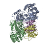
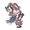
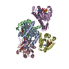
| Title | Bacillus subtilis Complex structure 1 of diadenylate cyclase CdaA cytoplasmic domain (CdaACD) and the phosphoglucomutase GlmM short variant (GlmMF369) | ||||||
 Components Components |
| ||||||
 Keywords Keywords | PROTEIN BINDING / Phosphoglucosamine Mutase / glucosamine-6-phosphate / glucosamine-1-phosphate / c-di-AMP / diadenylate cyclase / GlmM / CdaA / DacA / protein protein complex / isomerase / heterodimer | ||||||
| Function / homology |  Function and homology information Function and homology informationphosphoglucosamine mutase / phosphoglucosamine mutase activity / phosphomannomutase activity / diadenylate cyclase / diadenylate cyclase activity / UDP-N-acetylglucosamine biosynthetic process / adenylate cyclase activity / cAMP biosynthetic process / peptidoglycan biosynthetic process / carbohydrate metabolic process ...phosphoglucosamine mutase / phosphoglucosamine mutase activity / phosphomannomutase activity / diadenylate cyclase / diadenylate cyclase activity / UDP-N-acetylglucosamine biosynthetic process / adenylate cyclase activity / cAMP biosynthetic process / peptidoglycan biosynthetic process / carbohydrate metabolic process / magnesium ion binding / ATP binding / plasma membrane / cytosol Similarity search - Function | ||||||
| Biological species |  | ||||||
| Method |  X-RAY DIFFRACTION / SYNCHROTRON / MOLECULAR REPLACEMENT / Resolution: 3.65 Å X-RAY DIFFRACTION / SYNCHROTRON / MOLECULAR REPLACEMENT / Resolution: 3.65 Å | ||||||
 Authors Authors | Pathania, M. / Grundling, A.G. / Freemont, P. | ||||||
| Funding support |  United Kingdom, 1items United Kingdom, 1items
| ||||||
 Citation Citation | Journal: J.Biol.Chem. / Year: 2021 Title: Structural basis for the inhibition of the Bacillus subtilis c-di-AMP cyclase CdaA by the phosphoglucomutase GlmM. Authors: Pathania, M. / Tosi, T. / Millership, C. / Hoshiga, F. / Morgan, R.M.L. / Freemont, P.S. / Grundling, A. | ||||||
| History |
|
- Structure visualization
| Structure viewer | Molecule: MolmilJmol/JSmol |
|---|
- Downloads & links
-Download
| PDBx/mmCIF format | 7olh.cif.gz | 579.9 KB | Display | PDBx/mmCIF format |
|---|---|---|---|---|
| PDB format | pdb7olh.ent.gz | 470.2 KB | Display | PDB format |
| PDBx/mmJSON format | 7olh.json.gz | Tree view | PDBx/mmJSON format | |
| Others |  Other downloads Other downloads |
-Validation report
| Arichive directory | https://data.pdbj.org/pub/pdb/validation_reports/ol/7olhftp://data.pdbj.org/pub/pdb/validation_reports/ol/7olh | HTTPS FTP |
|---|
-Related structure data
| Related structure data | 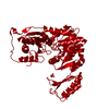 7ojrC 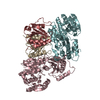 7ojsC  7omlC C: citing same article ( |
|---|---|
| Similar structure data | |
| Other databases |
|


-Links
 PDBj
PDBj- Assembly
| Deposited unit | 
| |||||||||||||||||||||||||||||||||
|---|---|---|---|---|---|---|---|---|---|---|---|---|---|---|---|---|---|---|---|---|---|---|---|---|---|---|---|---|---|---|---|---|---|---|
| 1 | 
| |||||||||||||||||||||||||||||||||
| 2 | 
| |||||||||||||||||||||||||||||||||
| 3 | 
| |||||||||||||||||||||||||||||||||
| Unit cell |
| |||||||||||||||||||||||||||||||||
| Noncrystallographic symmetry (NCS) | NCS domain:
|