 Movie
Movie Controller

[English] 日本語
 Yorodumi
- PDB-7ols: MerTK kinase domain with type 1.5 inhibitor containing a di-methy... -
+ Open data

- Basic information
| Entry | Database: PDB / ID: 7ols | ||||||
|---|---|---|---|---|---|---|---|
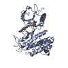
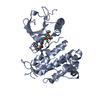
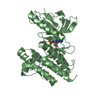
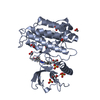
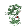
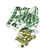
| Title | MerTK kinase domain with type 1.5 inhibitor containing a di-methyl pyrazole group | ||||||
 Components Components | Tyrosine-protein kinase Mer | ||||||
 Keywords Keywords | TRANSFERASE / tyrosine kinase inhibitor / structure-based drug design / type 1.5 inhibitor | ||||||
| Function / homology |  Function and homology information Function and homology informationnegative regulation of leukocyte apoptotic process / negative regulation of lymphocyte activation / neutrophil clearance / natural killer cell differentiation / secretion by cell / negative regulation of cytokine production / vagina development / photoreceptor outer segment / phagocytosis / transmembrane receptor protein tyrosine kinase activity ...negative regulation of leukocyte apoptotic process / negative regulation of lymphocyte activation / neutrophil clearance / natural killer cell differentiation / secretion by cell / negative regulation of cytokine production / vagina development / photoreceptor outer segment / phagocytosis / transmembrane receptor protein tyrosine kinase activity / substrate adhesion-dependent cell spreading / establishment of localization in cell / positive regulation of phagocytosis / cell surface receptor protein tyrosine kinase signaling pathway / Cell surface interactions at the vascular wall / receptor protein-tyrosine kinase / platelet activation / cell-cell signaling / nervous system development / cell migration / retina development in camera-type eye / spermatogenesis / protein phosphorylation / cell surface receptor signaling pathway / positive regulation of phosphatidylinositol 3-kinase/protein kinase B signal transduction / signaling receptor complex / : / ATP binding / plasma membrane / cytoplasm Similarity search - Function | ||||||
| Biological species |  Homo sapiens (human) Homo sapiens (human) | ||||||
| Method |  X-RAY DIFFRACTION / SYNCHROTRON / MOLECULAR REPLACEMENT / Resolution: 1.89 Å X-RAY DIFFRACTION / SYNCHROTRON / MOLECULAR REPLACEMENT / Resolution: 1.89 Å | ||||||
 Authors Authors | Pflug, A. / Schimpl, M. / McCoull, W. / Nissink, J.W.M. / Winter-Holt, J. | ||||||
 Citation Citation | Journal: J.Med.Chem. / Year: 2021 Title: Optimization of an Imidazo[1,2- a ]pyridine Series to Afford Highly Selective Type I1/2 Dual Mer/Axl Kinase Inhibitors with In Vivo Efficacy. Authors: McCoull, W. / Boyd, S. / Brown, M.R. / Coen, M. / Collingwood, O. / Davies, N.L. / Doherty, A. / Fairley, G. / Goldberg, K. / Hardaker, E. / He, G. / Hennessy, E.J. / Hopcroft, P. / Hodgson, ...Authors: McCoull, W. / Boyd, S. / Brown, M.R. / Coen, M. / Collingwood, O. / Davies, N.L. / Doherty, A. / Fairley, G. / Goldberg, K. / Hardaker, E. / He, G. / Hennessy, E.J. / Hopcroft, P. / Hodgson, G. / Jackson, A. / Jiang, X. / Karmokar, A. / Laine, A.L. / Lindsay, N. / Mao, Y. / Markandu, R. / McMurray, L. / McLean, N. / Mooney, L. / Musgrove, H. / Nissink, J.W.M. / Pflug, A. / Reddy, V.P. / Rawlins, P.B. / Rivers, E. / Schimpl, M. / Smith, G.F. / Tentarelli, S. / Travers, J. / Troup, R.I. / Walton, J. / Wang, C. / Wilkinson, S. / Williamson, B. / Winter-Holt, J. / Yang, D. / Zheng, Y. / Zhu, Q. / Smith, P.D. | ||||||
| History |
|
- Structure visualization
| Structure viewer | Molecule: MolmilJmol/JSmol |
|---|
- Downloads & links
-Download
| PDBx/mmCIF format | 7ols.cif.gz | 74 KB | Display | PDBx/mmCIF format |
|---|---|---|---|---|
| PDB format | pdb7ols.ent.gz | 51.9 KB | Display | PDB format |
| PDBx/mmJSON format | 7ols.json.gz | Tree view | PDBx/mmJSON format | |
| Others |  Other downloads Other downloads |
-Validation report
| Arichive directory | https://data.pdbj.org/pub/pdb/validation_reports/ol/7olsftp://data.pdbj.org/pub/pdb/validation_reports/ol/7ols | HTTPS FTP |
|---|
-Related structure data
| Related structure data |  7olvC 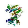 7olxC 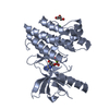 3brbS S: Starting model for refinement C: citing same article ( |
|---|---|
| Similar structure data |


-Links
 PDBj
PDBj


- Assembly
| Deposited unit | 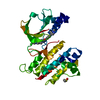
| ||||||||
|---|---|---|---|---|---|---|---|---|---|
| 1 |
| ||||||||
| Unit cell |
| ||||||||
| Components on special symmetry positions |
|
-Components
| #1: Protein | Mass: 34381.754 Da / Num. of mol.: 1 / Fragment: kinase domain (571-864) Source method: isolated from a genetically manipulated source Source: (gene. exp.) Homo sapiens (human) / Gene: MERTK, MER / Production host:  References: UniProt: Q12866, receptor protein-tyrosine kinase | ||||||
|---|---|---|---|---|---|---|---|
| #2: Chemical | ChemComp-VJZ /   Mass: 413.475 Da / Num. of mol.: 1 / Source method: obtained synthetically / Formula: C23H23N7O / Feature type: SUBJECT OF INVESTIGATION Mass: 413.475 Da / Num. of mol.: 1 / Source method: obtained synthetically / Formula: C23H23N7O / Feature type: SUBJECT OF INVESTIGATION | ||||||
| #3: Chemical | ChemComp-CL /   Mass: 35.453 Da / Num. of mol.: 4 / Source method: obtained synthetically / Formula: Cl Mass: 35.453 Da / Num. of mol.: 4 / Source method: obtained synthetically / Formula: Cl#4: Chemical | ChemComp-DMS / |   Mass: 78.133 Da / Num. of mol.: 1 / Source method: obtained synthetically / Formula: C2H6OS / Comment: DMSO, precipitant*YM Mass: 78.133 Da / Num. of mol.: 1 / Source method: obtained synthetically / Formula: C2H6OS / Comment: DMSO, precipitant*YM#5: Water | ChemComp-HOH / |  Mass: 18.015 Da / Num. of mol.: 107 / Source method: isolated from a natural source / Formula: H2O Mass: 18.015 Da / Num. of mol.: 107 / Source method: isolated from a natural source / Formula: H2OHas ligand of interest | Y | |
-Experimental details
-Experiment
| Experiment | Method: X-RAY DIFFRACTION / Number of used crystals: 1 |
|---|
- Sample preparation
| Crystal | Density Matthews: 2.27 Å3/Da / Density % sol: 45.9 % |
|---|---|
| Crystal grow | Temperature: 293 K / Method: vapor diffusion / Details: 0.1 M Tris pH 8.5, 4.3 M NaCl |
-Data collection
| Diffraction | Mean temperature: 100 K / Serial crystal experiment: N | |||||||||||||||||||||||||||
|---|---|---|---|---|---|---|---|---|---|---|---|---|---|---|---|---|---|---|---|---|---|---|---|---|---|---|---|---|
| Diffraction source | Source: SYNCHROTRON / Site: Diamond  / Beamline: I03 / Wavelength: 0.97625 Å / Beamline: I03 / Wavelength: 0.97625 Å | |||||||||||||||||||||||||||
| Detector | Type: DECTRIS PILATUS 6M / Detector: PIXEL / Date: Jul 7, 2017 | |||||||||||||||||||||||||||
| Radiation | Protocol: SINGLE WAVELENGTH / Monochromatic (M) / Laue (L): M / Scattering type: x-ray | |||||||||||||||||||||||||||
| Radiation wavelength | Wavelength: 0.97625 Å / Relative weight: 1 | |||||||||||||||||||||||||||
| Reflection | Resolution: 1.89→66.07 Å / Num. obs: 16521 / % possible obs: 94.6 % / Redundancy: 6.4 % / CC1/2: 0.999 / Rmerge(I) obs: 0.071 / Rpim(I) all: 0.031 / Rrim(I) all: 0.078 / Net I/σ(I): 15.3 | |||||||||||||||||||||||||||
| Reflection shell | Diffraction-ID: 1 / Redundancy: 5.9 %
|
- Processing
| Software |
| ||||||||||||||||||||||||||||||||||||||||||||||||||||||||||||
|---|---|---|---|---|---|---|---|---|---|---|---|---|---|---|---|---|---|---|---|---|---|---|---|---|---|---|---|---|---|---|---|---|---|---|---|---|---|---|---|---|---|---|---|---|---|---|---|---|---|---|---|---|---|---|---|---|---|---|---|---|---|
| Refinement | Method to determine structure: MOLECULAR REPLACEMENT Starting model: 3BRB Resolution: 1.89→66.07 Å / Cor.coef. Fo:Fc: 0.948 / Cor.coef. Fo:Fc free: 0.891 / SU B: 5.526 / SU ML: 0.153 / SU R Cruickshank DPI: 0.2871 / Cross valid method: THROUGHOUT / σ(F): 0 / ESU R: 0.287 / ESU R Free: 0.239 / Stereochemistry target values: MAXIMUM LIKELIHOOD Details: HYDROGENS HAVE BEEN ADDED IN THE RIDING POSITIONS U VALUES : REFINED INDIVIDUALLY
| ||||||||||||||||||||||||||||||||||||||||||||||||||||||||||||
| Solvent computation | Ion probe radii: 0.8 Å / Shrinkage radii: 0.8 Å / VDW probe radii: 1.2 Å / Solvent model: MASK | ||||||||||||||||||||||||||||||||||||||||||||||||||||||||||||
| Displacement parameters | Biso max: 95.66 Å2 / Biso mean: 47.483 Å2 / Biso min: 23.89 Å2
| ||||||||||||||||||||||||||||||||||||||||||||||||||||||||||||
| Refinement step | Cycle: final / Resolution: 1.89→66.07 Å
| ||||||||||||||||||||||||||||||||||||||||||||||||||||||||||||
| Refine LS restraints |
| ||||||||||||||||||||||||||||||||||||||||||||||||||||||||||||
| LS refinement shell | Resolution: 1.895→1.944 Å / Rfactor Rfree error: 0
|
