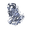
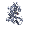
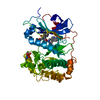
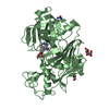
 Movie
Movie Controller


+ Open data

- Basic information
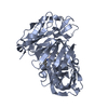
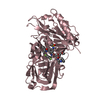
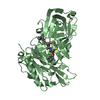
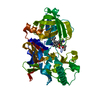
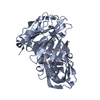
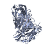
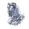
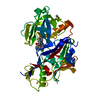
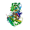
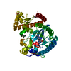
| Entry | Database: PDB / ID: 6lyj | ||||||||||||
|---|---|---|---|---|---|---|---|---|---|---|---|---|---|
| Title | The crystal structure of SAUGI/EBVUDG complex | ||||||||||||
 Components Components |
| ||||||||||||
 Keywords Keywords | HYDROLASE / DNA mimic protein / uracil-DNA glycosylase inhibitor / uracil-DNA glycosylase / DNA repair / herpesvirus | ||||||||||||
| Function / homology |  Function and homology information Function and homology informationbase-excision repair, AP site formation via deaminated base removal / uracil-DNA glycosylase / uracil DNA N-glycosylase activity / host cell nucleus / metal ion binding Similarity search - Function | ||||||||||||
| Biological species |  Epstein-Barr virus (Epstein-Barr virus) Epstein-Barr virus (Epstein-Barr virus)  Staphylococcus aureus (bacteria) Staphylococcus aureus (bacteria) | ||||||||||||
| Method |  X-RAY DIFFRACTION / SYNCHROTRON / MOLECULAR REPLACEMENT / Resolution: 2.1 Å X-RAY DIFFRACTION / SYNCHROTRON / MOLECULAR REPLACEMENT / Resolution: 2.1 Å | ||||||||||||
 Authors Authors | Liao, Y.T. / Ko, T.P. / Wang, H.C. | ||||||||||||
| Funding support |  Taiwan, 3items Taiwan, 3items
| ||||||||||||
 Citation Citation | Journal: Int.J.Biol.Macromol. / Year: 2020 Title: Structural insight into the differential interactions between the DNA mimic protein SAUGI and two gamma herpesvirus uracil-DNA glycosylases. Authors: Liao, Y.T. / Lin, S.J. / Ko, T.P. / Liu, C.Y. / Hsu, K.C. / Wang, H.C. | ||||||||||||
| History |
|
- Structure visualization
| Structure viewer | Molecule: MolmilJmol/JSmol |
|---|
- Downloads & links
-Download
| PDBx/mmCIF format | 6lyj.cif.gz | 357 KB | Display | PDBx/mmCIF format |
|---|---|---|---|---|
| PDB format | pdb6lyj.ent.gz | 241.1 KB | Display | PDB format |
| PDBx/mmJSON format | 6lyj.json.gz | Tree view | PDBx/mmJSON format | |
| Others |  Other downloads Other downloads |
-Validation report
| Arichive directory | https://data.pdbj.org/pub/pdb/validation_reports/ly/6lyjftp://data.pdbj.org/pub/pdb/validation_reports/ly/6lyj | HTTPS FTP |
|---|
-Related structure data
| Related structure data |  6lyvC  5aysS S: Starting model for refinement C: citing same article ( |
|---|---|
| Similar structure data |


-Links
 PDBj
PDBj- Assembly
| Deposited unit | 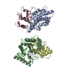
| |||||||||||||||||||||||||||||||||||||||||||||||||||||||||||||||||||||||||
|---|---|---|---|---|---|---|---|---|---|---|---|---|---|---|---|---|---|---|---|---|---|---|---|---|---|---|---|---|---|---|---|---|---|---|---|---|---|---|---|---|---|---|---|---|---|---|---|---|---|---|---|---|---|---|---|---|---|---|---|---|---|---|---|---|---|---|---|---|---|---|---|---|---|---|
| 1 | 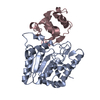
| |||||||||||||||||||||||||||||||||||||||||||||||||||||||||||||||||||||||||
| 2 | 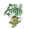
| |||||||||||||||||||||||||||||||||||||||||||||||||||||||||||||||||||||||||
| Unit cell |
| |||||||||||||||||||||||||||||||||||||||||||||||||||||||||||||||||||||||||
| Noncrystallographic symmetry (NCS) | NCS domain:
NCS domain segments:
NCS ensembles :
|
-Components
| #1: Protein | Mass: 28642.053 Da / Num. of mol.: 2 Source method: isolated from a genetically manipulated source Source: (gene. exp.) Epstein-Barr virus (strain GD1) (Epstein-Barr virus)Strain: GD1 Gene: BKRF3, EBVaGC1_032, HHV4_BKRF3, SNU-719_036, YCCEL1_037 Production host: References: UniProt: Q3KSS2, UniProt: P12888*PLUS, uracil-DNA glycosylase #2: Protein | Mass: 13367.123 Da / Num. of mol.: 2 Source method: isolated from a genetically manipulated source Source: (gene. exp.) Staphylococcus aureus (bacteria) / Production host: #3: Water | ChemComp-HOH / |  Mass: 18.015 Da / Num. of mol.: 758 / Source method: isolated from a natural source / Formula: H2O Mass: 18.015 Da / Num. of mol.: 758 / Source method: isolated from a natural source / Formula: H2O |
|---|
-Experimental details
-Experiment
| Experiment | Method: X-RAY DIFFRACTION / Number of used crystals: 1 |
|---|
- Sample preparation
| Crystal | Density Matthews: 2.16 Å3/Da / Density % sol: 43.07 % |
|---|---|
| Crystal grow | Temperature: 293 K / Method: evaporation / Details: TrisHCl, PEG 6000 |
-Data collection
| Diffraction | Mean temperature: 100 K / Serial crystal experiment: N |
|---|---|
| Diffraction source | Source: SYNCHROTRON / Site: NSRRC / Beamline: BL13C1 / Wavelength: 0.97622 Å |
| Detector | Type: ADSC QUANTUM 315r / Detector: CCD / Date: Jun 14, 2017 |
| Radiation | Protocol: SINGLE WAVELENGTH / Monochromatic (M) / Laue (L): M / Scattering type: x-ray |
| Radiation wavelength | Wavelength: 0.97622 Å / Relative weight: 1 |
| Reflection | Resolution: 2.1→20 Å / Num. obs: 41768 / % possible obs: 96.5 % / Redundancy: 5.1 % / Biso Wilson estimate: 25.18 Å2 / Rmerge(I) obs: 0.079 / Net I/σ(I): 27.7 |
| Reflection shell | Resolution: 2.1→2.17 Å / Rmerge(I) obs: 0.356 / Num. unique obs: 3865 |
- Processing
| Software |
| |||||||||||||||||||||||||||||||||||||||||||||||||||||||||||||||||||||||||||||||||||||||||||||||||||||||||||||||||||||||||||||
|---|---|---|---|---|---|---|---|---|---|---|---|---|---|---|---|---|---|---|---|---|---|---|---|---|---|---|---|---|---|---|---|---|---|---|---|---|---|---|---|---|---|---|---|---|---|---|---|---|---|---|---|---|---|---|---|---|---|---|---|---|---|---|---|---|---|---|---|---|---|---|---|---|---|---|---|---|---|---|---|---|---|---|---|---|---|---|---|---|---|---|---|---|---|---|---|---|---|---|---|---|---|---|---|---|---|---|---|---|---|---|---|---|---|---|---|---|---|---|---|---|---|---|---|---|---|---|
| Refinement | Method to determine structure: MOLECULAR REPLACEMENT Starting model: 5AYS Resolution: 2.1→19.95 Å / SU ML: 0.2188 / Cross valid method: FREE R-VALUE / σ(F): 1.34 / Phase error: 21.7221
| |||||||||||||||||||||||||||||||||||||||||||||||||||||||||||||||||||||||||||||||||||||||||||||||||||||||||||||||||||||||||||||
| Solvent computation | Shrinkage radii: 0.9 Å / VDW probe radii: 1.11 Å | |||||||||||||||||||||||||||||||||||||||||||||||||||||||||||||||||||||||||||||||||||||||||||||||||||||||||||||||||||||||||||||
| Displacement parameters | Biso mean: 32.36 Å2 | |||||||||||||||||||||||||||||||||||||||||||||||||||||||||||||||||||||||||||||||||||||||||||||||||||||||||||||||||||||||||||||
| Refinement step | Cycle: LAST / Resolution: 2.1→19.95 Å
| |||||||||||||||||||||||||||||||||||||||||||||||||||||||||||||||||||||||||||||||||||||||||||||||||||||||||||||||||||||||||||||
| Refine LS restraints |
| |||||||||||||||||||||||||||||||||||||||||||||||||||||||||||||||||||||||||||||||||||||||||||||||||||||||||||||||||||||||||||||
| LS refinement shell |
| |||||||||||||||||||||||||||||||||||||||||||||||||||||||||||||||||||||||||||||||||||||||||||||||||||||||||||||||||||||||||||||
| Refinement TLS params. | Method: refined / Refine-ID: X-RAY DIFFRACTION
| |||||||||||||||||||||||||||||||||||||||||||||||||||||||||||||||||||||||||||||||||||||||||||||||||||||||||||||||||||||||||||||
| Refinement TLS group |
|
