 Movie
Movie Controller

[English] 日本語
 Yorodumi
- PDB-7ojy: Crystal structure of Pseudomonas aeruginosa LpxA in complex with ... -
+ Open data

- Basic information
| Entry | Database: PDB / ID: 7ojy | ||||||
|---|---|---|---|---|---|---|---|
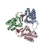
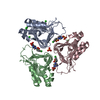
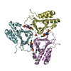
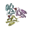
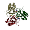
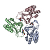
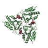
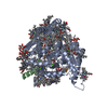
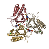
| Title | Crystal structure of Pseudomonas aeruginosa LpxA in complex with compound 6 | ||||||
 Components Components | Acyl-[acyl-carrier-protein]-UDP-N-acetylglucosamine O-acyltransferase | ||||||
 Keywords Keywords | TRANSFERASE / ACYLTRANSFERASE / FATTY ACIDS / LIPID A / LEFT-HANDED PARALLEL BETA-HELIX | ||||||
| Function / homology |  Function and homology information Function and homology informationacyl-[acyl-carrier-protein]-UDP-N-acetylglucosamine O-acyltransferase / acyl-[acyl-carrier-protein]-UDP-N-acetylglucosamine O-acyltransferase activity / lipid A biosynthetic process / membrane / cytoplasm Similarity search - Function | ||||||
| Biological species |   Pseudomonas aeruginosa (bacteria) Pseudomonas aeruginosa (bacteria) | ||||||
| Method |  X-RAY DIFFRACTION / SYNCHROTRON / MOLECULAR REPLACEMENT / Resolution: 2 Å X-RAY DIFFRACTION / SYNCHROTRON / MOLECULAR REPLACEMENT / Resolution: 2 Å | ||||||
 Authors Authors | Ryan, M.D. / Parkes, A.L. / Southey, M. / Andersen, O.A. / Zahn, M. / Barker, J. / DeJonge, B.L.M. | ||||||
 Citation Citation | Journal: J.Med.Chem. / Year: 2021 Title: Discovery of Novel UDP- N -Acetylglucosamine Acyltransferase (LpxA) Inhibitors with Activity against Pseudomonas aeruginosa . Authors: Ryan, M.D. / Parkes, A.L. / Corbett, D. / Dickie, A.P. / Southey, M. / Andersen, O.A. / Stein, D.B. / Barbeau, O.R. / Sanzone, A. / Thommes, P. / Barker, J. / Cain, R. / Compper, C. / Dejob, ...Authors: Ryan, M.D. / Parkes, A.L. / Corbett, D. / Dickie, A.P. / Southey, M. / Andersen, O.A. / Stein, D.B. / Barbeau, O.R. / Sanzone, A. / Thommes, P. / Barker, J. / Cain, R. / Compper, C. / Dejob, M. / Dorali, A. / Etheridge, D. / Evans, S. / Faulkner, A. / Gadouleau, E. / Gorman, T. / Haase, D. / Holbrow-Wilshaw, M. / Krulle, T. / Li, X. / Lumley, C. / Mertins, B. / Napier, S. / Odedra, R. / Papadopoulos, K. / Roumpelakis, V. / Spear, K. / Trimby, E. / Williams, J. / Zahn, M. / Keefe, A.D. / Zhang, Y. / Soutter, H.T. / Centrella, P.A. / Clark, M.A. / Cuozzo, J.W. / Dumelin, C.E. / Deng, B. / Hunt, A. / Sigel, E.A. / Troast, D.M. / DeJonge, B.L.M. | ||||||
| History |
|
- Structure visualization
| Structure viewer | Molecule: MolmilJmol/JSmol |
|---|
- Downloads & links
-Download
| PDBx/mmCIF format | 7ojy.cif.gz | 320.4 KB | Display | PDBx/mmCIF format |
|---|---|---|---|---|
| PDB format | pdb7ojy.ent.gz | 259.9 KB | Display | PDB format |
| PDBx/mmJSON format | 7ojy.json.gz | Tree view | PDBx/mmJSON format | |
| Others |  Other downloads Other downloads |
-Validation report
| Arichive directory | https://data.pdbj.org/pub/pdb/validation_reports/oj/7ojyftp://data.pdbj.org/pub/pdb/validation_reports/oj/7ojy | HTTPS FTP |
|---|
-Related structure data
| Related structure data | 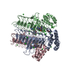 7oj6C 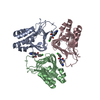 7ojpC 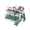 7ojqC 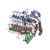 7ojwC 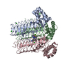 7ok1C 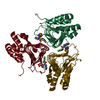 7ok2C 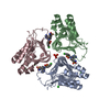 7okaC  7okbC 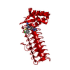 7okcC  5demS S: Starting model for refinement C: citing same article ( |
|---|---|
| Similar structure data |

-Links
 PDBj
PDBj


- Assembly
| Deposited unit | 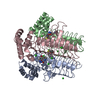
| ||||||||
|---|---|---|---|---|---|---|---|---|---|
| 1 |
| ||||||||
| Unit cell |
|
-Components
| #1: Protein | Mass: 28331.010 Da / Num. of mol.: 3 / Fragment: full-length protein Source method: isolated from a genetically manipulated source Source: (gene. exp.) Pseudomonas aeruginosa (strain PA7) (bacteria)Strain: PA7 / Gene: lpxA, PSPA7_1495 / Plasmid: pET28b / Production host: References: UniProt: A6V1E4, acyl-[acyl-carrier-protein]-UDP-N-acetylglucosamine O-acyltransferase #2: Chemical |   Mass: 414.865 Da / Num. of mol.: 3 / Source method: obtained synthetically / Formula: C19H15ClN4O3S / Feature type: SUBJECT OF INVESTIGATION Mass: 414.865 Da / Num. of mol.: 3 / Source method: obtained synthetically / Formula: C19H15ClN4O3S / Feature type: SUBJECT OF INVESTIGATION#3: Chemical | ChemComp-CL /   Mass: 35.453 Da / Num. of mol.: 14 / Source method: obtained synthetically / Formula: Cl Mass: 35.453 Da / Num. of mol.: 14 / Source method: obtained synthetically / Formula: Cl#4: Water | ChemComp-HOH / |  Mass: 18.015 Da / Num. of mol.: 513 / Source method: isolated from a natural source / Formula: H2O Mass: 18.015 Da / Num. of mol.: 513 / Source method: isolated from a natural source / Formula: H2OHas ligand of interest | Y | |
|---|
-Experimental details
-Experiment
| Experiment | Method: X-RAY DIFFRACTION / Number of used crystals: 1 |
|---|
- Sample preparation
| Crystal | Density Matthews: 4.05 Å3/Da / Density % sol: 69.6 % |
|---|---|
| Crystal grow | Temperature: 278 K / Method: vapor diffusion, hanging drop / pH: 4.5 / Details: 1.5 M NH4Cl, 0.1 M Na Acetate pH 4.5 |
-Data collection
| Diffraction | Mean temperature: 100 K / Serial crystal experiment: N | ||||||||||||||||||||||||
|---|---|---|---|---|---|---|---|---|---|---|---|---|---|---|---|---|---|---|---|---|---|---|---|---|---|
| Diffraction source | Source: SYNCHROTRON / Site: Diamond  / Beamline: I04 / Wavelength: 0.9795 Å / Beamline: I04 / Wavelength: 0.9795 Å | ||||||||||||||||||||||||
| Detector | Type: DECTRIS EIGER X 16M / Detector: PIXEL / Date: Apr 12, 2019 | ||||||||||||||||||||||||
| Radiation | Protocol: SINGLE WAVELENGTH / Monochromatic (M) / Laue (L): M / Scattering type: x-ray | ||||||||||||||||||||||||
| Radiation wavelength | Wavelength: 0.9795 Å / Relative weight: 1 | ||||||||||||||||||||||||
| Reflection | Resolution: 2→83.2 Å / Num. obs: 91413 / % possible obs: 99.7 % / Redundancy: 6.9 % / Biso Wilson estimate: 32.87 Å2 / CC1/2: 0.99 / Rmerge(I) obs: 0.162 / Rpim(I) all: 0.067 / Rrim(I) all: 0.175 / Net I/σ(I): 4.7 | ||||||||||||||||||||||||
| Reflection shell | Diffraction-ID: 1
|
- Processing
| Software |
| ||||||||||||||||||||||||||||||||||||||||||||||||||||||||||||||||||||||||||||||||||||||||||||||||||||||||||||
|---|---|---|---|---|---|---|---|---|---|---|---|---|---|---|---|---|---|---|---|---|---|---|---|---|---|---|---|---|---|---|---|---|---|---|---|---|---|---|---|---|---|---|---|---|---|---|---|---|---|---|---|---|---|---|---|---|---|---|---|---|---|---|---|---|---|---|---|---|---|---|---|---|---|---|---|---|---|---|---|---|---|---|---|---|---|---|---|---|---|---|---|---|---|---|---|---|---|---|---|---|---|---|---|---|---|---|---|---|---|
| Refinement | Method to determine structure: MOLECULAR REPLACEMENT Starting model: 5DEM Resolution: 2→58.83 Å / Cor.coef. Fo:Fc: 0.938 / Cor.coef. Fo:Fc free: 0.915 / SU R Cruickshank DPI: 0.122 / Cross valid method: THROUGHOUT / σ(F): 0 / SU R Blow DPI: 0.124 / SU Rfree Blow DPI: 0.118 / SU Rfree Cruickshank DPI: 0.117
| ||||||||||||||||||||||||||||||||||||||||||||||||||||||||||||||||||||||||||||||||||||||||||||||||||||||||||||
| Displacement parameters | Biso max: 133.03 Å2 / Biso mean: 48.35 Å2 / Biso min: 26.29 Å2
| ||||||||||||||||||||||||||||||||||||||||||||||||||||||||||||||||||||||||||||||||||||||||||||||||||||||||||||
| Refine analyze | Luzzati coordinate error obs: 0.26 Å | ||||||||||||||||||||||||||||||||||||||||||||||||||||||||||||||||||||||||||||||||||||||||||||||||||||||||||||
| Refinement step | Cycle: final / Resolution: 2→58.83 Å
| ||||||||||||||||||||||||||||||||||||||||||||||||||||||||||||||||||||||||||||||||||||||||||||||||||||||||||||
| Refine LS restraints |
| ||||||||||||||||||||||||||||||||||||||||||||||||||||||||||||||||||||||||||||||||||||||||||||||||||||||||||||
| LS refinement shell | Resolution: 2→2.01 Å / Rfactor Rfree error: 0 / Total num. of bins used: 50
| ||||||||||||||||||||||||||||||||||||||||||||||||||||||||||||||||||||||||||||||||||||||||||||||||||||||||||||
| Refinement TLS params. | Method: refined / Refine-ID: X-RAY DIFFRACTION
| ||||||||||||||||||||||||||||||||||||||||||||||||||||||||||||||||||||||||||||||||||||||||||||||||||||||||||||
| Refinement TLS group |
|
