 ムービー
ムービー コントローラー


+ データを開く

- 基本情報
基本情報
| 登録情報 | データベース: PDB / ID: 7ohf | |||||||||
|---|---|---|---|---|---|---|---|---|---|---|
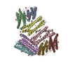
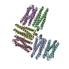
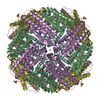
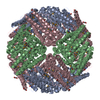
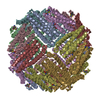
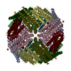
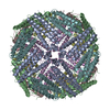
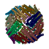
| タイトル | Cryo-EM structure of pyrococcus furiosus apoferritin in nanofluidic channels | |||||||||
 要素 要素 | Ferritin | |||||||||
 キーワード キーワード | METAL TRANSPORT / Iron Storage | |||||||||
| 機能・相同性 |  機能・相同性情報 機能・相同性情報ferroxidase activity / ferric iron binding / iron ion transport / ferrous iron binding / intracellular iron ion homeostasis / identical protein binding / cytosol 類似検索 - 分子機能 | |||||||||
| 生物種 |   Pyrococcus furiosus COM1 (古細菌) Pyrococcus furiosus COM1 (古細菌) | |||||||||
| 手法 | 電子顕微鏡法 / 単粒子再構成法 / クライオ電子顕微鏡法 / 解像度: 3 Å | |||||||||
 データ登録者 データ登録者 | Huber, S.T. / Sarajlic, E. / Huijink, R. / Evers, W.H. / Jakobi, A.J. | |||||||||
| 資金援助 | European Union,  オランダ, 2件 オランダ, 2件
| |||||||||
 引用 引用 | ジャーナル: Elife / 年: 2022 タイトル: Nanofluidic chips for cryo-EM structure determination from picoliter sample volumes. 著者: Stefan T Huber / Edin Sarajlic / Roeland Huijink / Felix Weis / Wiel H Evers / Arjen J Jakobi /  要旨: Cryogenic electron microscopy has become an essential tool for structure determination of biological macromolecules. In practice, the difficulty to reliably prepare samples with uniform ice thickness ...Cryogenic electron microscopy has become an essential tool for structure determination of biological macromolecules. In practice, the difficulty to reliably prepare samples with uniform ice thickness still represents a barrier for routine high-resolution imaging and limits the current throughput of the technique. We show that a nanofluidic sample support with well-defined geometry can be used to prepare cryo-EM specimens with reproducible ice thickness from picoliter sample volumes. The sample solution is contained in electron-transparent nanochannels that provide uniform thickness gradients without further optimisation and eliminate the potentially destructive air-water interface. We demonstrate the possibility to perform high-resolution structure determination with three standard protein specimens. Nanofabricated sample supports bear potential to automate the cryo-EM workflow, and to explore new frontiers for cryo-EM applications such as time-resolved imaging and high-throughput screening. #1: ジャーナル: Biorxiv / 年: 2021タイトル: Nanofluidic chips for cryo-EM structure determination from picoliter sample volumes 著者: Huber, S.T. / Sarajlic, E. / Huijink, R. / Weis, F. / Evers, W.H. / Jakobi, A.J. | |||||||||
| 履歴 |
|
- 構造の表示
構造の表示
| ムービー |
ムービービューア |
|---|---|
| 構造ビューア | 分子: MolmilJmol/JSmol |
- ダウンロードとリンク
-ダウンロード
| PDBx/mmCIF形式 | 7ohf.cif.gz | 42.7 KB | 表示 | PDBx/mmCIF形式 |
|---|---|---|---|---|
| PDB形式 | pdb7ohf.ent.gz | 28.6 KB | 表示 | PDB形式 |
| PDBx/mmJSON形式 | 7ohf.json.gz | ツリー表示 | PDBx/mmJSON形式 | |
| その他 |  その他のダウンロード その他のダウンロード |
-検証レポート
| 文書・要旨 | 7ohf_validation.pdf.gz | 1.5 MB | 表示 | wwPDB検証レポート |
|---|---|---|---|---|
| 文書・詳細版 | 7ohf_full_validation.pdf.gz | 1.5 MB | 表示 | |
| XML形式データ | 7ohf_validation.xml.gz | 25 KB | 表示 | |
| CIF形式データ | 7ohf_validation.cif.gz | 33.2 KB | 表示 | |
| アーカイブディレクトリ | https://data.pdbj.org/pub/pdb/validation_reports/oh/7ohfftp://data.pdbj.org/pub/pdb/validation_reports/oh/7ohf | HTTPS FTP |
-関連構造データ
| 関連構造データ |  12901MC M: このデータのモデリングに利用したマップデータ C: 同じ文献を引用 ( |
|---|---|
| 類似構造データ | |
| 電子顕微鏡画像生データ | EMPIAR-10708 (タイトル: Apoferritin, TMV and T20S proteasome in nanofluidic channels Data size: 308.3 Data #1: Unaligned multi-frame movies of pyrococcus furiosus apoferritin in silicon nitride nanochannels [micrographs - multiframe] Data #2: Multi-frame movies of T20S proteasome in silicon nitride nanochannels [micrographs - multiframe] Data #3: Multi-frame movies of TMV in silicon nitride nanochannels [micrographs - multiframe]) |


-リンク
 PDBj
PDBj


- 集合体
集合体
| 登録構造単位 | 
|
|---|---|
| 1 | x 24
|
-要素
| #1: タンパク質 | 分子量: 20535.395 Da / 分子数: 1 / 由来タイプ: 組換発現 / 由来: (組換発現) Pyrococcus furiosus COM1 (古細菌) / 遺伝子: PFC_02870 / 発現宿主:  |
|---|
-実験情報
-実験
| 実験 | 手法: 電子顕微鏡法 |
|---|---|
| EM実験 | 試料の集合状態: PARTICLE / 3次元再構成法: 単粒子再構成法 |
- 試料調製
試料調製
| 構成要素 | 名称: 24-mer of pyrococcus furiosus apoferritin / タイプ: COMPLEX / Entity ID: all / 由来: RECOMBINANT | |||||||||||||||
|---|---|---|---|---|---|---|---|---|---|---|---|---|---|---|---|---|
| 分子量 | 値: 0.492 MDa / 実験値: NO | |||||||||||||||
| 由来(天然) | 生物種: Pyrococcus furiosus COM1 (古細菌) | |||||||||||||||
| 由来(組換発現) | 生物種: | |||||||||||||||
| 緩衝液 | pH: 7.5 | |||||||||||||||
| 緩衝液成分 |
| |||||||||||||||
| 試料 | 濃度: 3.4 mg/ml / 包埋: NO / シャドウイング: NO / 染色: NO / 凍結: YES / 詳細: The sample was filled into nanofluidic channels. | |||||||||||||||
| 試料支持 | グリッドの材料: SILICON NITRIDE / グリッドのタイプ: Homemade | |||||||||||||||
| 急速凍結 | 装置: LEICA PLUNGER / 凍結剤: ETHANE 詳細: The sample was filled into cryoChips through the cantilever and then transferred within ~10 seconds to the Leica plunger for freezing. |
- 電子顕微鏡撮影
| 実験機器 |  モデル: Titan Krios / 画像提供: FEI Company |
|---|---|
| 顕微鏡 | モデル: FEI TITAN KRIOS |
| 電子銃 | 電子線源:  FIELD EMISSION GUN / 加速電圧: 300 kV / 照射モード: FLOOD BEAM FIELD EMISSION GUN / 加速電圧: 300 kV / 照射モード: FLOOD BEAM |
| 電子レンズ | モード: BRIGHT FIELD / 最大 デフォーカス(公称値): -2000 nm / 最小 デフォーカス(公称値): -1000 nm / Cs: 2.7 mm |
| 試料ホルダ | 試料ホルダーモデル: FEI TITAN KRIOS AUTOGRID HOLDER |
| 撮影 | 平均露光時間: 9 sec. / 電子線照射量: 63 e/Å2 / 検出モード: COUNTING フィルム・検出器のモデル: GATAN K2 SUMMIT (4k x 4k) 撮影したグリッド数: 3 / 実像数: 948 |
| 画像スキャン | 動画フレーム数/画像: 90 |
- 解析
解析
| ソフトウェア | 名称: PHENIX / バージョン: 1.13_2998: / 分類: 精密化 | ||||||||||||||||||||||||||||||||||||
|---|---|---|---|---|---|---|---|---|---|---|---|---|---|---|---|---|---|---|---|---|---|---|---|---|---|---|---|---|---|---|---|---|---|---|---|---|---|
| EMソフトウェア |
| ||||||||||||||||||||||||||||||||||||
| CTF補正 | タイプ: PHASE FLIPPING AND AMPLITUDE CORRECTION | ||||||||||||||||||||||||||||||||||||
| 3次元再構成 | 解像度: 3 Å / 解像度の算出法: FSC 0.143 CUT-OFF / 粒子像の数: 21238 / クラス平均像の数: 1 / 対称性のタイプ: POINT | ||||||||||||||||||||||||||||||||||||
| 原子モデル構築 | PDB-ID: 2X17 Accession code: 2X17 / Source name: PDB / タイプ: experimental model |

