 Movie
Movie Controller

[English] 日本語
 Yorodumi
- EMDB-12902: Electron cryo-tomogram of pyrococcus furiosus apoferritin in nano... -
+ Open data

- Basic information
| Entry | Database: EMDB / ID: EMD-12902 | |||||||||
|---|---|---|---|---|---|---|---|---|---|---|
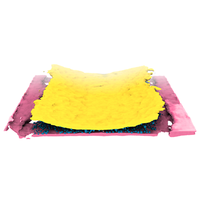
| Title | Electron cryo-tomogram of pyrococcus furiosus apoferritin in nanofluidic channels | |||||||||
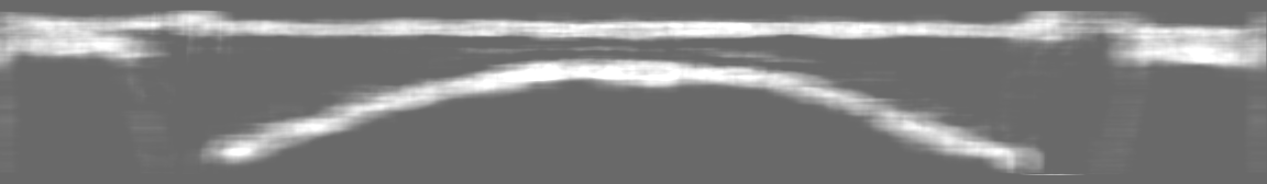
 Map data Map data | Tomogram of nanofluidic channels filled with 3.4 mg/mL apoferritin solution | |||||||||
 Sample Sample |
| |||||||||
| Biological species |   Pyrococcus furiosus COM1 (archaea) Pyrococcus furiosus COM1 (archaea) | |||||||||
| Method | electron tomography / cryo EM | |||||||||
 Authors Authors | Huber ST / Sarajlic E / Huijink R / Evers WH / Jakobi AJ | |||||||||
| Funding support | European Union,  Netherlands, 2 items Netherlands, 2 items
| |||||||||
 Citation Citation | Journal: Elife / Year: 2022 Title: Nanofluidic chips for cryo-EM structure determination from picoliter sample volumes. Authors: Stefan T Huber / Edin Sarajlic / Roeland Huijink / Felix Weis / Wiel H Evers / Arjen J Jakobi /  Abstract: Cryogenic electron microscopy has become an essential tool for structure determination of biological macromolecules. In practice, the difficulty to reliably prepare samples with uniform ice thickness ...Cryogenic electron microscopy has become an essential tool for structure determination of biological macromolecules. In practice, the difficulty to reliably prepare samples with uniform ice thickness still represents a barrier for routine high-resolution imaging and limits the current throughput of the technique. We show that a nanofluidic sample support with well-defined geometry can be used to prepare cryo-EM specimens with reproducible ice thickness from picoliter sample volumes. The sample solution is contained in electron-transparent nanochannels that provide uniform thickness gradients without further optimisation and eliminate the potentially destructive air-water interface. We demonstrate the possibility to perform high-resolution structure determination with three standard protein specimens. Nanofabricated sample supports bear potential to automate the cryo-EM workflow, and to explore new frontiers for cryo-EM applications such as time-resolved imaging and high-throughput screening. #1: Journal: Biorxiv / Year: 2021Title: Nanofluidic chips for cryo-EM structure determination from picoliter sample volumes Authors: Huber ST / Sarajlic E / Huijink R / Weis F / Evers WH / Jakobi AJ | |||||||||
| History |
|
- Structure visualization
| Movie |
 Movie viewer Movie viewer |
|---|---|
| Supplemental images |
- Downloads & links
-EMDB archive
| Map data | emd_12902.map.gz | 1.1 GB | EMDB map data format | |
|---|---|---|---|---|
| Header (meta data) | emd-12902-v30.xmlemd-12902.xml | 14.3 KB 14.3 KB | Display Display | EMDB header |
| Images |  emd_12902.png emd_12902.png | 62.2 KB | ||
| Others | emd_12902_additional_1.map.gzemd_12902_additional_2.map.gz | 273 MB 605.1 MB | ||
| Archive directory |  http://ftp.pdbj.org/pub/emdb/structures/EMD-12902ftp://ftp.pdbj.org/pub/emdb/structures/EMD-12902 http://ftp.pdbj.org/pub/emdb/structures/EMD-12902ftp://ftp.pdbj.org/pub/emdb/structures/EMD-12902 | HTTPS FTP |
-Related structure data


-Links
| EMDB pages | EMDB (EBI/PDBe) / EMDataResource |
|---|
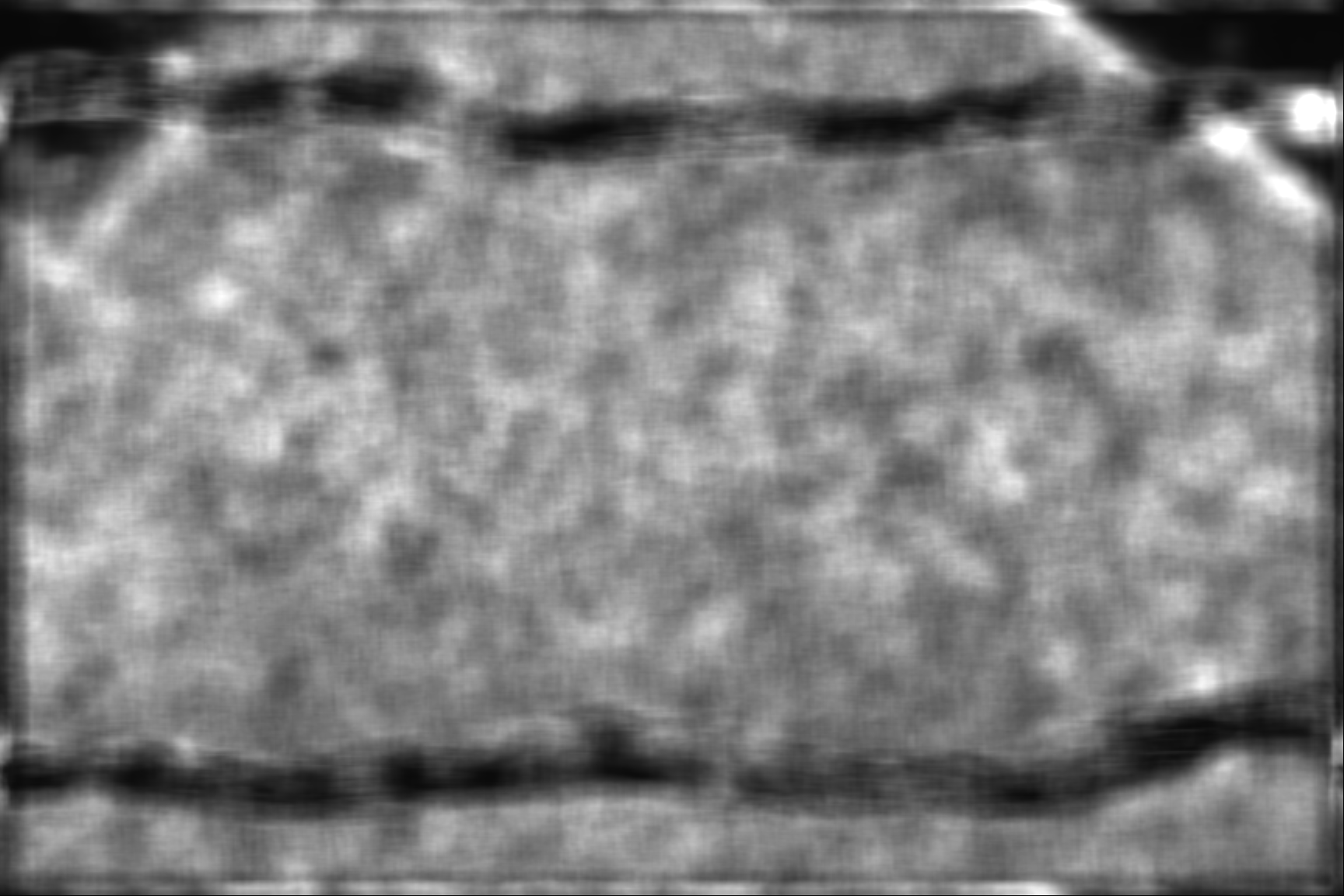
-Map
| File | Download / File: emd_12902.map.gz / Format: CCP4 / Size: 1.2 GB / Type: IMAGE STORED AS FLOATING POINT NUMBER (4 BYTES) | ||||||||||||||||||||||||||||||||||||||||||||||||||||||||||||
|---|---|---|---|---|---|---|---|---|---|---|---|---|---|---|---|---|---|---|---|---|---|---|---|---|---|---|---|---|---|---|---|---|---|---|---|---|---|---|---|---|---|---|---|---|---|---|---|---|---|---|---|---|---|---|---|---|---|---|---|---|---|
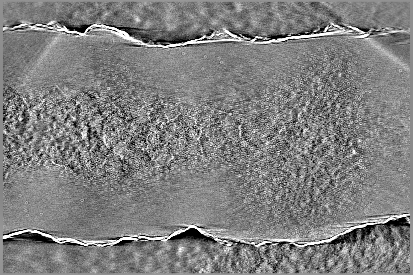
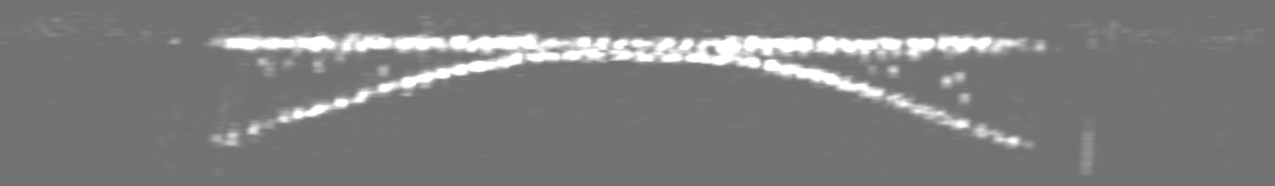
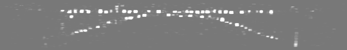
| Annotation | Tomogram of nanofluidic channels filled with 3.4 mg/mL apoferritin solution | ||||||||||||||||||||||||||||||||||||||||||||||||||||||||||||
| Projections & slices | Image control
Images are generated by Spider. generated in cubic-lattice coordinate | ||||||||||||||||||||||||||||||||||||||||||||||||||||||||||||
| Voxel size | X=Y=Z: 9.412 Å | ||||||||||||||||||||||||||||||||||||||||||||||||||||||||||||
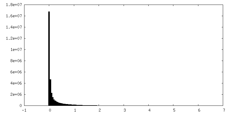
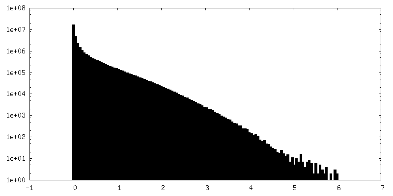
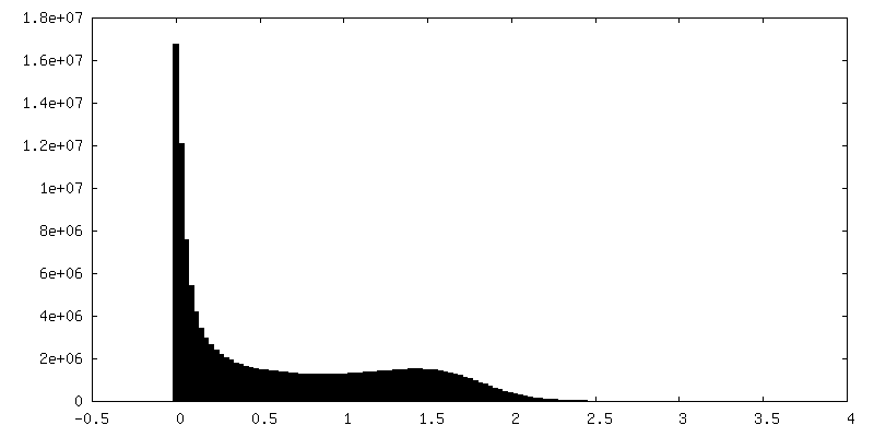
| Density |
| ||||||||||||||||||||||||||||||||||||||||||||||||||||||||||||
| Symmetry | Space group: 1 | ||||||||||||||||||||||||||||||||||||||||||||||||||||||||||||
| Details | EMDB XML:
CCP4 map header:
| ||||||||||||||||||||||||||||||||||||||||||||||||||||||||||||
 Z (Sec.)
 Y (Row.)
 X (Col.)


-Supplemental data
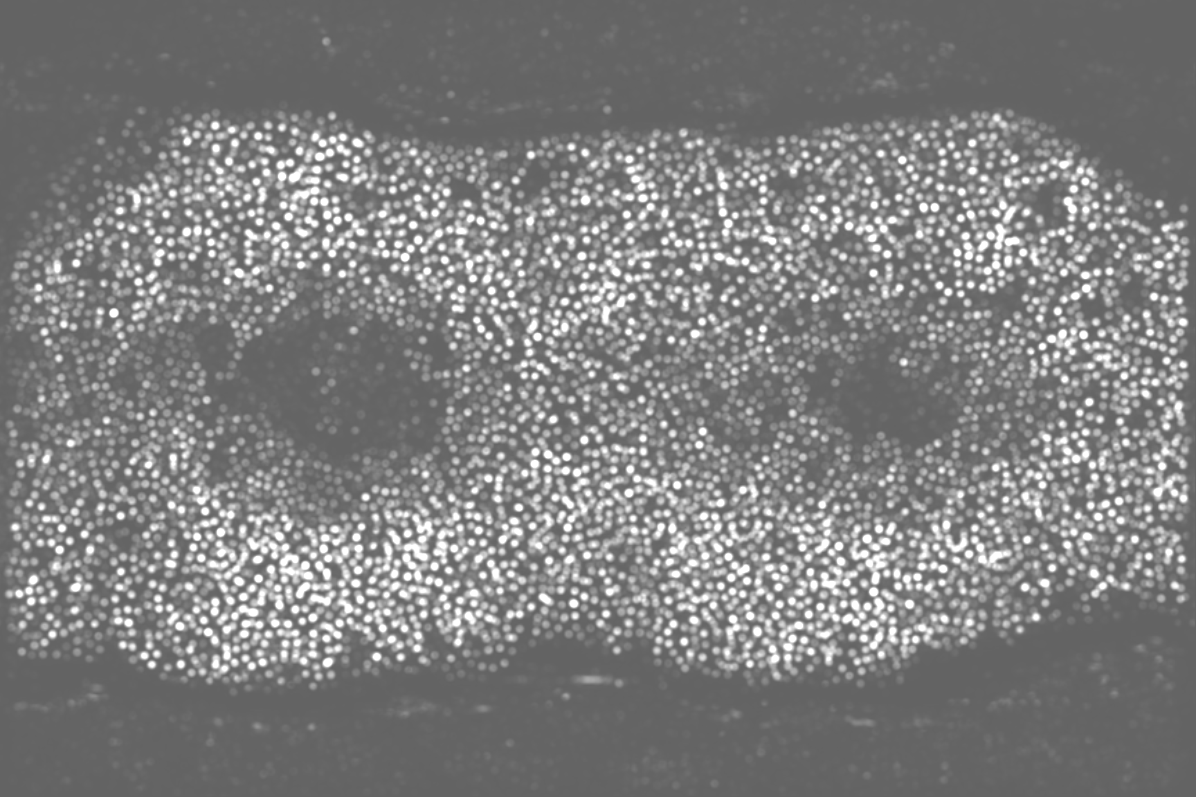
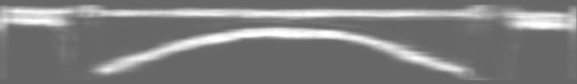
-Additional map: Segmentation of apoferritin particles
| File | emd_12902_additional_1.map | ||||||||||||
|---|---|---|---|---|---|---|---|---|---|---|---|---|---|
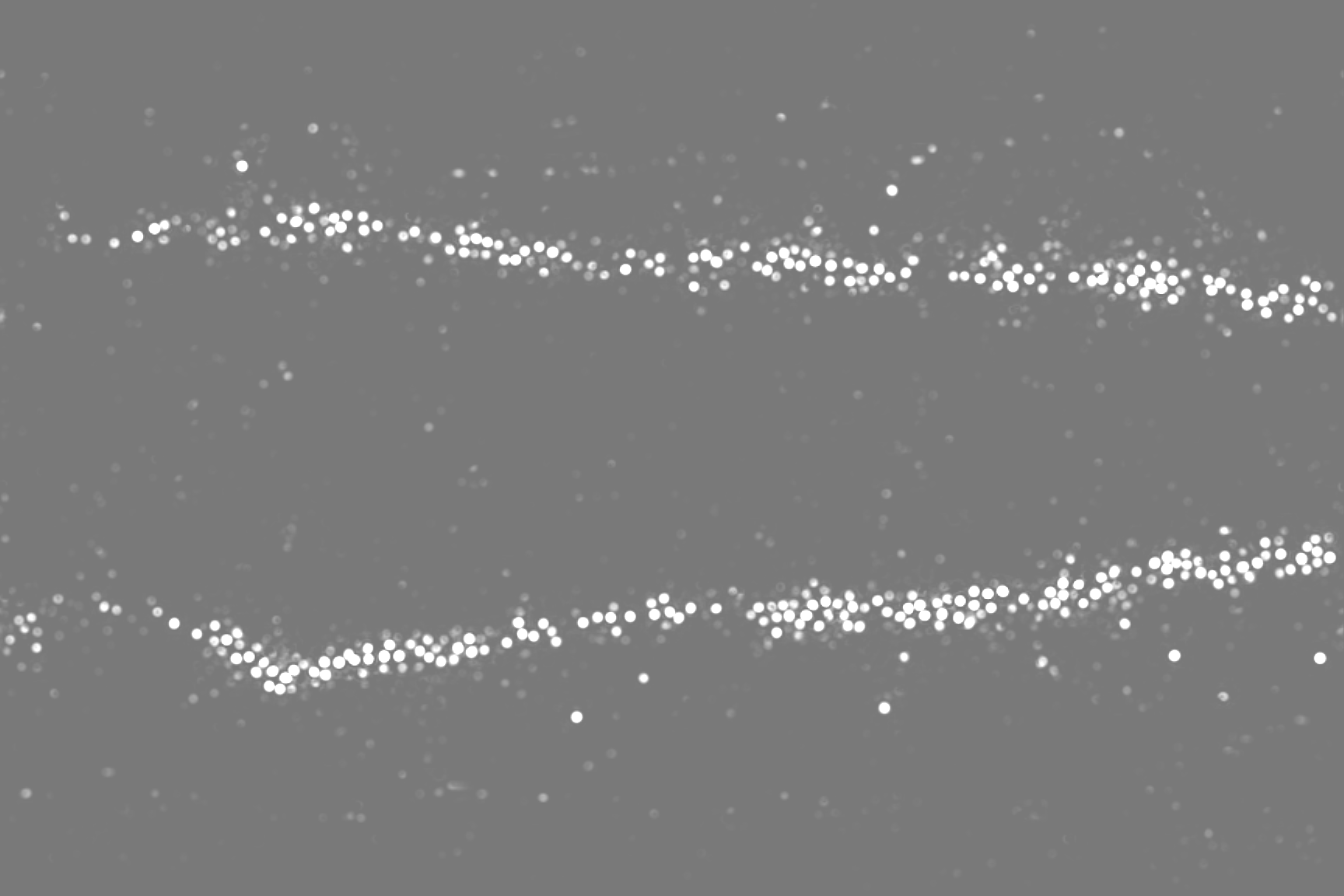
| Annotation | Segmentation of apoferritin particles | ||||||||||||
| Projections & Slices |
| ||||||||||||
| Density Histograms |


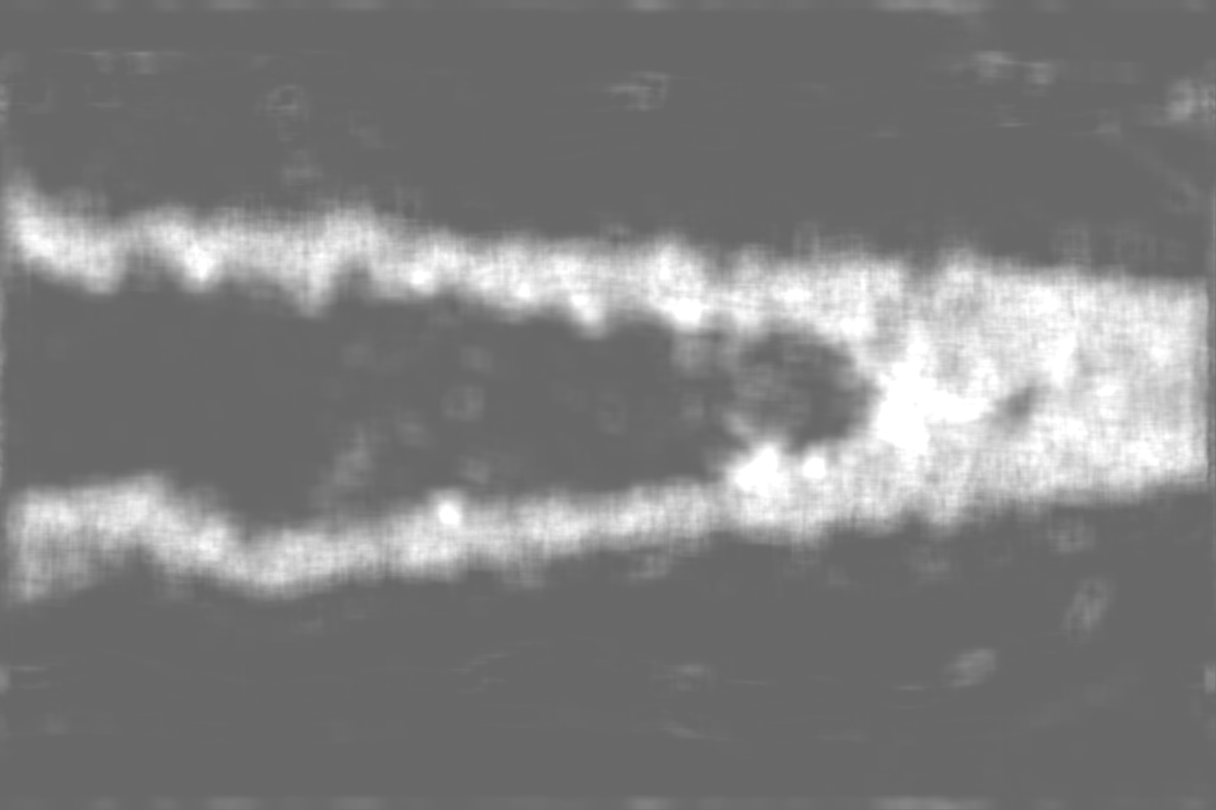
-Additional map: Segmentation of the silicon nitride membrane
| File | emd_12902_additional_2.map | ||||||||||||
|---|---|---|---|---|---|---|---|---|---|---|---|---|---|
| Annotation | Segmentation of the silicon nitride membrane | ||||||||||||
| Projections & Slices |
| ||||||||||||
| Density Histograms |


- Sample components
-Entire : Nanochannel filled with pyrococcus furiosus apoferritin
| Entire | Name: Nanochannel filled with pyrococcus furiosus apoferritin |
|---|---|
| Components |
|
-Supramolecule #1: Nanochannel filled with pyrococcus furiosus apoferritin
| Supramolecule | Name: Nanochannel filled with pyrococcus furiosus apoferritin type: complex / ID: 1 / Parent: 0 |
|---|---|
| Source (natural) | Organism: Pyrococcus furiosus COM1 (archaea) |
| Recombinant expression | Organism:  |
| Molecular weight | Theoretical: 492 KDa |
-Experimental details
-Structure determination
| Method | cryo EM |
|---|---|
 Processing Processing | electron tomography |
| Aggregation state | particle |
-Sample preparation
| Concentration | 3.4 mg/mL | |||||||||
|---|---|---|---|---|---|---|---|---|---|---|
| Buffer | pH: 7.5 Component:
| |||||||||
| Grid | Model: Homemade / Material: SILICON NITRIDE | |||||||||
| Vitrification | Cryogen name: ETHANE / Instrument: LEICA PLUNGER Details: cryoChips were filled with 3.4 mg/mL apoferritin and transferred to the Leica plunger within ~10 seconds for plunging.. | |||||||||
| Sectioning | Other: NO SECTIONING |
- Electron microscopy
| Microscope | JEOL 3200FSC |
|---|---|
| Image recording | Film or detector model: GATAN K2 SUMMIT (4k x 4k) / Detector mode: COUNTING / Average electron dose: 94.6 e/Å2 |
| Electron beam | Acceleration voltage: 300 kV / Electron source:  FIELD EMISSION GUN FIELD EMISSION GUN |
| Electron optics | Illumination mode: FLOOD BEAM / Imaging mode: BRIGHT FIELD / Cs: 4.1 mm / Nominal defocus min: 6.0 µm |
-Image processing
| Final reconstruction | Algorithm: BACK PROJECTION / Resolution method: OTHER / Number images used: 61 |
|---|---|
| CTF correction | Software - Name: IMOD (ver. 4.9.2) |
