 Movie
Movie Controller


+ Open data

- Basic information
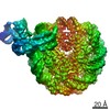
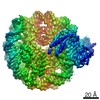
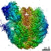
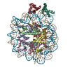
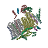
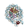
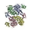
| Entry | Database: PDB / ID: 7ohb | ||||||||||||||||||||||||||||||||||||
|---|---|---|---|---|---|---|---|---|---|---|---|---|---|---|---|---|---|---|---|---|---|---|---|---|---|---|---|---|---|---|---|---|---|---|---|---|---|
| Title | TBP-nucleosome complex | ||||||||||||||||||||||||||||||||||||
 Components Components |
| ||||||||||||||||||||||||||||||||||||
 Keywords Keywords | TRANSCRIPTION / DNA binding protein nucleosome | ||||||||||||||||||||||||||||||||||||
| Function / homology |  Function and homology information Function and homology informationtranscription initiation at RNA polymerase II promoter / structural constituent of chromatin / nucleosome / nucleosome assembly / heterochromatin formation / transcription regulator complex / protein heterodimerization activity / DNA binding / nucleoplasm / nucleus Similarity search - Function | ||||||||||||||||||||||||||||||||||||
| Biological species | synthetic construct (others)  | ||||||||||||||||||||||||||||||||||||
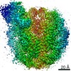
| Method | ELECTRON MICROSCOPY / single particle reconstruction / cryo EM / Resolution: 3.4 Å | ||||||||||||||||||||||||||||||||||||
 Authors Authors | Wang, H. / Cramer, P. | ||||||||||||||||||||||||||||||||||||
| Funding support |  Germany, European Union, 2items Germany, European Union, 2items
| ||||||||||||||||||||||||||||||||||||
 Citation Citation | Journal: Proc Natl Acad Sci U S A / Year: 2021 Title: Structures and implications of TBP-nucleosome complexes. Authors: Haibo Wang / Le Xiong / Patrick Cramer / Abstract: The TATA box-binding protein (TBP) is highly conserved throughout eukaryotes and plays a central role in the assembly of the transcription preinitiation complex (PIC) at gene promoters. TBP binds and ...The TATA box-binding protein (TBP) is highly conserved throughout eukaryotes and plays a central role in the assembly of the transcription preinitiation complex (PIC) at gene promoters. TBP binds and bends DNA, and directs adjacent binding of the transcription factors TFIIA and TFIIB for PIC assembly. Here, we show that yeast TBP can bind to a nucleosome containing the Widom-601 sequence and that TBP-nucleosome binding is stabilized by TFIIA. We determine three cryo-electron microscopy (cryo-EM) structures of TBP-nucleosome complexes, two of them containing also TFIIA. TBP can bind to superhelical location (SHL) -6, which contains a TATA-like sequence, but also to SHL +2, which is GC-rich. Whereas binding to SHL -6 can occur in the absence of TFIIA, binding to SHL +2 is only observed in the presence of TFIIA and goes along with detachment of upstream terminal DNA from the histone octamer. TBP-nucleosome complexes are sterically incompatible with PIC assembly, explaining why a promoter nucleosome generally impairs transcription and must be moved before initiation can occur. | ||||||||||||||||||||||||||||||||||||
| History |
|
- Structure visualization
| Movie |
Movie viewer |
|---|---|
| Structure viewer | Molecule: MolmilJmol/JSmol |
- Downloads & links
-Download
| PDBx/mmCIF format | 7ohb.cif.gz | 316.5 KB | Display | PDBx/mmCIF format |
|---|---|---|---|---|
| PDB format | pdb7ohb.ent.gz | 239.3 KB | Display | PDB format |
| PDBx/mmJSON format | 7ohb.json.gz | Tree view | PDBx/mmJSON format | |
| Others |  Other downloads Other downloads |
-Validation report
| Arichive directory | https://data.pdbj.org/pub/pdb/validation_reports/oh/7ohbftp://data.pdbj.org/pub/pdb/validation_reports/oh/7ohb | HTTPS FTP |
|---|
-Related structure data
| Related structure data | 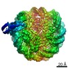 12899MC 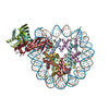 7oh9C 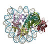 7ohaC 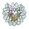 7ohcC M: map data used to model this data C: citing same article ( |
|---|---|
| Similar structure data |

-Links
 PDBj
PDBj


- Assembly
| Deposited unit | 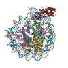
|
|---|---|
| 1 |
|
-Components
-Protein , 5 types, 9 molecules AEBFCGDHK
| #1: Protein | Mass: 15303.930 Da / Num. of mol.: 2 Source method: isolated from a genetically manipulated source Source: (gene. exp.)  #2: Protein | Mass: 11263.231 Da / Num. of mol.: 2 Source method: isolated from a genetically manipulated source Source: (gene. exp.) #3: Protein | Mass: 13978.241 Da / Num. of mol.: 2 Source method: isolated from a genetically manipulated source Source: (gene. exp.) #4: Protein | Mass: 13524.752 Da / Num. of mol.: 2 Source method: isolated from a genetically manipulated source Source: (gene. exp.) #7: Protein | | Mass: 27042.275 Da / Num. of mol.: 1 Source method: isolated from a genetically manipulated source Source: (gene. exp.) Gene: SPT15 / Production host: |
|---|
-DNA chain , 2 types, 2 molecules IJ
| #5: DNA chain | Mass: 44520.383 Da / Num. of mol.: 1 Source method: isolated from a genetically manipulated source Source: (gene. exp.) synthetic construct (others) / Production host: |
|---|---|
| #6: DNA chain | Mass: 44991.660 Da / Num. of mol.: 1 Source method: isolated from a genetically manipulated source Source: (gene. exp.) synthetic construct (others) / Production host: |
-Details
| Has protein modification | N |
|---|
-Experimental details
-Experiment
| Experiment | Method: ELECTRON MICROSCOPY |
|---|---|
| EM experiment | Aggregation state: PARTICLE / 3D reconstruction method: single particle reconstruction |
- Sample preparation
| Component | Name: Nucleosome with TBP / Type: COMPLEX / Entity ID: all / Source: MULTIPLE SOURCES |
|---|---|
| Molecular weight | Value: 0.23 MDa / Experimental value: YES |
| Buffer solution | pH: 7.5 |
| Specimen | Conc.: 0.12 mg/ml / Embedding applied: NO / Shadowing applied: NO / Staining applied: NO / Vitrification applied: YES |
| Vitrification | Instrument: FEI VITROBOT MARK IV / Cryogen name: ETHANE / Humidity: 100 % / Chamber temperature: 277 K |
- Electron microscopy imaging
| Experimental equipment |  Model: Titan Krios / Image courtesy: FEI Company |
|---|---|
| Microscopy | Model: FEI TITAN KRIOS |
| Electron gun | Electron source:  FIELD EMISSION GUN / Accelerating voltage: 300 kV / Illumination mode: FLOOD BEAM FIELD EMISSION GUN / Accelerating voltage: 300 kV / Illumination mode: FLOOD BEAM |
| Electron lens | Mode: BRIGHT FIELD / Cs: 2.7 mm / C2 aperture diameter: 70 µm |
| Image recording | Electron dose: 41.2 e/Å2 / Film or detector model: GATAN K3 (6k x 4k) |
- Processing
| Software | Name: PHENIX / Version: 1.19.2_4158: / Classification: refinement | ||||||||||||||||||||||||
|---|---|---|---|---|---|---|---|---|---|---|---|---|---|---|---|---|---|---|---|---|---|---|---|---|---|
| EM software | Name: PHENIX / Category: model refinement | ||||||||||||||||||||||||
| CTF correction | Type: PHASE FLIPPING AND AMPLITUDE CORRECTION | ||||||||||||||||||||||||
| 3D reconstruction | Resolution: 3.4 Å / Resolution method: FSC 0.143 CUT-OFF / Num. of particles: 36781 / Symmetry type: POINT | ||||||||||||||||||||||||
| Atomic model building | Protocol: RIGID BODY FIT / Space: REAL | ||||||||||||||||||||||||
| Refine LS restraints |
|
