 Movie
Movie Controller

[English] 日本語
 Yorodumi
- PDB-7og7: Crystal structure of the copper chaperone NosL from Shewanella de... -
+ Open data

- Basic information
| Entry | Database: PDB / ID: 7og7 | |||||||||
|---|---|---|---|---|---|---|---|---|---|---|
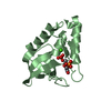
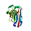
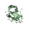
| Title | Crystal structure of the copper chaperone NosL from Shewanella denitrificans | |||||||||
 Components Components | NosL | |||||||||
 Keywords Keywords | METAL BINDING PROTEIN / copper chaperone | |||||||||
| Function / homology | Nitrous oxide reductase accessory protein NosL / NosL / metal ion binding / ACETONITRILE / COPPER (I) ION / NosL Function and homology information Function and homology information | |||||||||
| Biological species |  Shewanella denitrificans (bacteria) Shewanella denitrificans (bacteria) | |||||||||
| Method |  X-RAY DIFFRACTION / SYNCHROTRON / SAD / Resolution: 1.85 Å X-RAY DIFFRACTION / SYNCHROTRON / SAD / Resolution: 1.85 Å | |||||||||
 Authors Authors | Prasser, B. / Schoener, L. / Zhang, L. / Einsle, O. | |||||||||
| Funding support |  Germany, 2items Germany, 2items
| |||||||||
 Citation Citation | Journal: Angew.Chem.Int.Ed.Engl. / Year: 2021 Title: The Copper Chaperone NosL Forms a Heterometal Site for Cu Delivery to Nitrous Oxide Reductase. Authors: Prasser, B. / Schoner, L. / Zhang, L. / Einsle, O. | |||||||||
| History |
|
- Structure visualization
| Structure viewer | Molecule: MolmilJmol/JSmol |
|---|
- Downloads & links
-Download
| PDBx/mmCIF format | 7og7.cif.gz | 46.5 KB | Display | PDBx/mmCIF format |
|---|---|---|---|---|
| PDB format | pdb7og7.ent.gz | 32.3 KB | Display | PDB format |
| PDBx/mmJSON format | 7og7.json.gz | Tree view | PDBx/mmJSON format | |
| Others |  Other downloads Other downloads |
-Validation report
| Arichive directory | https://data.pdbj.org/pub/pdb/validation_reports/og/7og7ftp://data.pdbj.org/pub/pdb/validation_reports/og/7og7 | HTTPS FTP |
|---|
-Related structure data
| Similar structure data |
|---|


-Links
 PDBj
PDBj- Assembly
| Deposited unit | 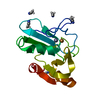
| ||||||||||||
|---|---|---|---|---|---|---|---|---|---|---|---|---|---|
| 1 |
| ||||||||||||
| Unit cell |
| ||||||||||||
| Components on special symmetry positions |
|
-Components
| #1: Protein | Mass: 20320.088 Da / Num. of mol.: 1 Source method: isolated from a genetically manipulated source Source: (gene. exp.) Shewanella denitrificans (strain OS217 / ATCC BAA-1090 / DSM 15013) (bacteria)Strain: OS217 / ATCC BAA-1090 / DSM 15013 / Gene: Sden_2215 / Production host: | ||||
|---|---|---|---|---|---|
| #2: Chemical | ChemComp-CU1 /   Mass: 63.546 Da / Num. of mol.: 1 / Source method: obtained synthetically / Formula: Cu / Feature type: SUBJECT OF INVESTIGATION Mass: 63.546 Da / Num. of mol.: 1 / Source method: obtained synthetically / Formula: Cu / Feature type: SUBJECT OF INVESTIGATION | ||||
| #3: Chemical | ChemComp-ZN /   Mass: 65.409 Da / Num. of mol.: 1 / Source method: obtained synthetically / Formula: Zn / Feature type: SUBJECT OF INVESTIGATION Mass: 65.409 Da / Num. of mol.: 1 / Source method: obtained synthetically / Formula: Zn / Feature type: SUBJECT OF INVESTIGATION | ||||
| #4: Chemical | ChemComp-CCN /   Mass: 41.052 Da / Num. of mol.: 5 / Source method: obtained synthetically / Formula: C2H3N Mass: 41.052 Da / Num. of mol.: 5 / Source method: obtained synthetically / Formula: C2H3N#5: Water | ChemComp-HOH / |  Mass: 18.015 Da / Num. of mol.: 102 / Source method: isolated from a natural source / Formula: H2O Mass: 18.015 Da / Num. of mol.: 102 / Source method: isolated from a natural source / Formula: H2OHas ligand of interest | Y | |
-Experimental details
-Experiment
| Experiment | Method: X-RAY DIFFRACTION / Number of used crystals: 1 |
|---|
- Sample preparation
| Crystal | Density Matthews: 2.94 Å3/Da / Density % sol: 58.21 % |
|---|---|
| Crystal grow | Temperature: 293 K / Method: vapor diffusion, sitting drop Details: 0.1 M HEPES buffer at pH 7.5 and 1.4 M trisodium citrate tribasic dihydrate |
-Data collection
| Diffraction | Mean temperature: 100 K / Serial crystal experiment: N |
|---|---|
| Diffraction source | Source: SYNCHROTRON / Site: SLS  / Beamline: X06SA / Wavelength: 1.28096 Å / Beamline: X06SA / Wavelength: 1.28096 Å |
| Detector | Type: DECTRIS EIGER X 16M / Detector: PIXEL / Date: Feb 8, 2016 |
| Radiation | Protocol: SINGLE WAVELENGTH / Monochromatic (M) / Laue (L): M / Scattering type: x-ray |
| Radiation wavelength | Wavelength: 1.28096 Å / Relative weight: 1 |
| Reflection | Resolution: 1.85→44.77 Å / Num. obs: 21586 / % possible obs: 100 % / Redundancy: 76.6 % / CC1/2: 1 / Net I/σ(I): 35.3 |
| Reflection shell | Resolution: 1.85→1.89 Å / Num. unique obs: 1322 / CC1/2: 0.431 |
- Processing
| Software |
| |||||||||||||||||||||||||||||||||||||||||||||||||||||||||||||||||||||||||||
|---|---|---|---|---|---|---|---|---|---|---|---|---|---|---|---|---|---|---|---|---|---|---|---|---|---|---|---|---|---|---|---|---|---|---|---|---|---|---|---|---|---|---|---|---|---|---|---|---|---|---|---|---|---|---|---|---|---|---|---|---|---|---|---|---|---|---|---|---|---|---|---|---|---|---|---|---|
| Refinement | Method to determine structure: SAD / Resolution: 1.85→44.767 Å / SU ML: 0.21 / Cross valid method: THROUGHOUT / σ(F): 2.02 / Phase error: 20.63 / Stereochemistry target values: ML
| |||||||||||||||||||||||||||||||||||||||||||||||||||||||||||||||||||||||||||
| Solvent computation | Shrinkage radii: 0.9 Å / VDW probe radii: 1.11 Å / Solvent model: FLAT BULK SOLVENT MODEL | |||||||||||||||||||||||||||||||||||||||||||||||||||||||||||||||||||||||||||
| Displacement parameters | Biso max: 77.79 Å2 / Biso mean: 35.9209 Å2 / Biso min: 20.35 Å2 | |||||||||||||||||||||||||||||||||||||||||||||||||||||||||||||||||||||||||||
| Refinement step | Cycle: final / Resolution: 1.85→44.767 Å
| |||||||||||||||||||||||||||||||||||||||||||||||||||||||||||||||||||||||||||
| LS refinement shell | Refine-ID: X-RAY DIFFRACTION / Rfactor Rfree error: 0 / % reflection obs: 100 %
|
