 Movie
Movie Controller

[English] 日本語
 Yorodumi
- PDB-7odx: Cyanophage S-2L Succinoaminodeoxyadenylate synthetase (PurZ) boun... -
+ Open data

- Basic information
| Entry | Database: PDB / ID: 7odx | ||||||
|---|---|---|---|---|---|---|---|
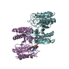
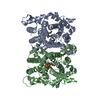
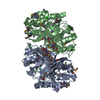
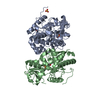
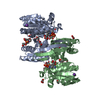
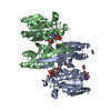
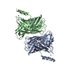
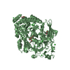
| Title | Cyanophage S-2L Succinoaminodeoxyadenylate synthetase (PurZ) bound to dGMP and dATP as an energy donor | ||||||
 Components Components | Succinoaminodeoxyadenylate synthetase (PurZ) | ||||||
 Keywords Keywords | VIRAL PROTEIN / S-2L / Succinoaminodeoxyadenylate synthetase / PurZ | ||||||
| Function / homology | 2'-DEOXYGUANOSINE-5'-MONOPHOSPHATE / 2'-DEOXYADENOSINE 5'-TRIPHOSPHATE Function and homology information Function and homology information | ||||||
| Biological species |  Cyanophage S-2L (virus) Cyanophage S-2L (virus) | ||||||
| Method |  X-RAY DIFFRACTION / SYNCHROTRON / MOLECULAR REPLACEMENT / Resolution: 1.69617086092 Å X-RAY DIFFRACTION / SYNCHROTRON / MOLECULAR REPLACEMENT / Resolution: 1.69617086092 Å | ||||||
 Authors Authors | Czernecki, D. / Delarue, M. | ||||||
 Citation Citation | Journal: Nat Commun / Year: 2021 Title: Characterization of a triad of genes in cyanophage S-2L sufficient to replace adenine by 2-aminoadenine in bacterial DNA. Authors: Czernecki, D. / Bonhomme, F. / Kaminski, P.A. / Delarue, M. | ||||||
| History |
|
- Structure visualization
| Structure viewer | Molecule: MolmilJmol/JSmol |
|---|
- Downloads & links
-Download
| PDBx/mmCIF format | 7odx.cif.gz | 190 KB | Display | PDBx/mmCIF format |
|---|---|---|---|---|
| PDB format | pdb7odx.ent.gz | 131.6 KB | Display | PDB format |
| PDBx/mmJSON format | 7odx.json.gz | Tree view | PDBx/mmJSON format | |
| Others |  Other downloads Other downloads |
-Validation report
| Arichive directory | https://data.pdbj.org/pub/pdb/validation_reports/od/7odxftp://data.pdbj.org/pub/pdb/validation_reports/od/7odx | HTTPS FTP |
|---|
-Related structure data
| Related structure data |  7odyC  6fm1S S: Starting model for refinement C: citing same article ( |
|---|---|
| Similar structure data |


-Links
 PDBj
PDBj


- Assembly
| Deposited unit | 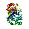
| ||||||||||||
|---|---|---|---|---|---|---|---|---|---|---|---|---|---|
| 1 | 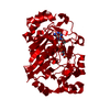
| ||||||||||||
| Unit cell |
| ||||||||||||
| Components on special symmetry positions |
|
-Components
| #1: Protein | Mass: 39897.113 Da / Num. of mol.: 1 Source method: isolated from a genetically manipulated source Source: (gene. exp.) Cyanophage S-2L (virus) / Production host:  |
|---|---|
| #2: Chemical | ChemComp-DGP /   Mass: 347.221 Da / Num. of mol.: 1 / Source method: obtained synthetically / Formula: C10H14N5O7P / Feature type: SUBJECT OF INVESTIGATION Mass: 347.221 Da / Num. of mol.: 1 / Source method: obtained synthetically / Formula: C10H14N5O7P / Feature type: SUBJECT OF INVESTIGATION |
| #3: Chemical | ChemComp-DTP /   Mass: 491.182 Da / Num. of mol.: 1 / Source method: obtained synthetically / Formula: C10H16N5O12P3 / Feature type: SUBJECT OF INVESTIGATION Mass: 491.182 Da / Num. of mol.: 1 / Source method: obtained synthetically / Formula: C10H16N5O12P3 / Feature type: SUBJECT OF INVESTIGATION |
| #4: Water | ChemComp-HOH /  Mass: 18.015 Da / Num. of mol.: 394 / Source method: isolated from a natural source / Formula: H2O Mass: 18.015 Da / Num. of mol.: 394 / Source method: isolated from a natural source / Formula: H2O |
| Has ligand of interest | Y |
-Experimental details
-Experiment
| Experiment | Method: X-RAY DIFFRACTION / Number of used crystals: 1 |
|---|
- Sample preparation
| Crystal | Density Matthews: 3.01 Å3/Da / Density % sol: 59.18 % |
|---|---|
| Crystal grow | Temperature: 277.15 K / Method: vapor diffusion, hanging drop / pH: 7 Details: 15% v/v Tacsimate; 2% w/v PEG 3350; 100 mM HEPES pH 7 |
-Data collection
| Diffraction | Mean temperature: 100 K / Serial crystal experiment: N |
|---|---|
| Diffraction source | Source: SYNCHROTRON / Site: SOLEIL  / Beamline: PROXIMA 2 / Wavelength: 0.98013 Å / Beamline: PROXIMA 2 / Wavelength: 0.98013 Å |
| Detector | Type: DECTRIS EIGER X 9M / Detector: PIXEL / Date: Sep 14, 2019 / Details: KB Mirrors |
| Radiation | Monochromator: Si 111 / Protocol: SINGLE WAVELENGTH / Monochromatic (M) / Laue (L): M / Scattering type: x-ray |
| Radiation wavelength | Wavelength: 0.98013 Å / Relative weight: 1 |
| Reflection | Resolution: 1.69617086092→44.5 Å / Num. obs: 54836 / % possible obs: 99.8 % / Redundancy: 39.6 % / Biso Wilson estimate: 28.6904003452 Å2 / CC1/2: 1 / Rmerge(I) obs: 0.081 / Rpim(I) all: 0.013 / Rrim(I) all: 0.082 / Net I/σ(I): 34.1 |
| Reflection shell | Resolution: 1.7→1.74 Å / Redundancy: 39.4 % / Rmerge(I) obs: 2.661 / Mean I/σ(I) obs: 1.7 / Num. unique obs: 3903 / CC1/2: 0.694 / Rpim(I) all: 0.419 / Rrim(I) all: 2.695 / % possible all: 98 |
- Processing
| Software |
| |||||||||||||||||||||||||||||||||||||||||||||||||||||||||||||||||||||||||||||||||||||||||||||||||||||||||||||||||||||||||||||||||||||||||||||||||||
|---|---|---|---|---|---|---|---|---|---|---|---|---|---|---|---|---|---|---|---|---|---|---|---|---|---|---|---|---|---|---|---|---|---|---|---|---|---|---|---|---|---|---|---|---|---|---|---|---|---|---|---|---|---|---|---|---|---|---|---|---|---|---|---|---|---|---|---|---|---|---|---|---|---|---|---|---|---|---|---|---|---|---|---|---|---|---|---|---|---|---|---|---|---|---|---|---|---|---|---|---|---|---|---|---|---|---|---|---|---|---|---|---|---|---|---|---|---|---|---|---|---|---|---|---|---|---|---|---|---|---|---|---|---|---|---|---|---|---|---|---|---|---|---|---|---|---|---|---|
| Refinement | Method to determine structure: MOLECULAR REPLACEMENT Starting model: 6FM1 Resolution: 1.69617086092→44.4954227156 Å / SU ML: 0.258644893723 / Cross valid method: FREE R-VALUE / σ(F): 1.33794450571 / Phase error: 17.7125464286 Stereochemistry target values: GeoStd + Monomer Library + CDL v1.2
| |||||||||||||||||||||||||||||||||||||||||||||||||||||||||||||||||||||||||||||||||||||||||||||||||||||||||||||||||||||||||||||||||||||||||||||||||||
| Solvent computation | Shrinkage radii: 0.9 Å / VDW probe radii: 1.11 Å / Solvent model: FLAT BULK SOLVENT MODEL | |||||||||||||||||||||||||||||||||||||||||||||||||||||||||||||||||||||||||||||||||||||||||||||||||||||||||||||||||||||||||||||||||||||||||||||||||||
| Displacement parameters | Biso mean: 32.2959699757 Å2 | |||||||||||||||||||||||||||||||||||||||||||||||||||||||||||||||||||||||||||||||||||||||||||||||||||||||||||||||||||||||||||||||||||||||||||||||||||
| Refinement step | Cycle: LAST / Resolution: 1.69617086092→44.4954227156 Å
| |||||||||||||||||||||||||||||||||||||||||||||||||||||||||||||||||||||||||||||||||||||||||||||||||||||||||||||||||||||||||||||||||||||||||||||||||||
| Refine LS restraints |
| |||||||||||||||||||||||||||||||||||||||||||||||||||||||||||||||||||||||||||||||||||||||||||||||||||||||||||||||||||||||||||||||||||||||||||||||||||
| LS refinement shell |
| |||||||||||||||||||||||||||||||||||||||||||||||||||||||||||||||||||||||||||||||||||||||||||||||||||||||||||||||||||||||||||||||||||||||||||||||||||
| Refinement TLS params. | Method: refined / Origin x: -38.8667272725 Å / Origin y: 22.2396766755 Å / Origin z: 4.48387363243 Å
| |||||||||||||||||||||||||||||||||||||||||||||||||||||||||||||||||||||||||||||||||||||||||||||||||||||||||||||||||||||||||||||||||||||||||||||||||||
| Refinement TLS group | Selection details: all |
