 Movie
Movie Controller

[English] 日本語
 Yorodumi
- PDB-7ocm: K1K1H6, a potent recombinant minimal hepatocyte growth factor/sca... -
+ Open data

- Basic information
| Entry | Database: PDB / ID: 7ocm | ||||||
|---|---|---|---|---|---|---|---|
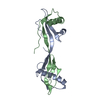
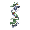
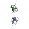
| Title | K1K1H6, a potent recombinant minimal hepatocyte growth factor/scatter factor mimic | ||||||
 Components Components | Hepatocyte growth factor alpha chain,Hepatocyte growth factor alpha chain | ||||||
 Keywords Keywords | DE NOVO PROTEIN / MET receptor agonist / HGF/SF kringle 1 dimer / HGF/SF-derived recombinant protein / MET-activator / regeneration of epithelial tissue and organs / engineered growth factor | ||||||
| Function / homology |  Function and homology information Function and homology informationregulation of p38MAPK cascade / skeletal muscle cell proliferation / regulation of branching involved in salivary gland morphogenesis by mesenchymal-epithelial signaling / Drug-mediated inhibition of MET activation / MET activates STAT3 / negative regulation of hydrogen peroxide-mediated programmed cell death / MET Receptor Activation / MET interacts with TNS proteins / MET receptor recycling / MET activates PTPN11 ...regulation of p38MAPK cascade / skeletal muscle cell proliferation / regulation of branching involved in salivary gland morphogenesis by mesenchymal-epithelial signaling / Drug-mediated inhibition of MET activation / MET activates STAT3 / negative regulation of hydrogen peroxide-mediated programmed cell death / MET Receptor Activation / MET interacts with TNS proteins / MET receptor recycling / MET activates PTPN11 / hepatocyte growth factor receptor signaling pathway / MET activates RAP1 and RAC1 / MET activates PI3K/AKT signaling / MET activates PTK2 signaling / cellular response to hepatocyte growth factor stimulus / positive regulation of DNA biosynthetic process / negative regulation of release of cytochrome c from mitochondria / chemoattractant activity / negative regulation of interleukin-6 production / myoblast proliferation / positive regulation of interleukin-10 production / epithelial to mesenchymal transition / positive regulation of osteoblast differentiation / negative regulation of extrinsic apoptotic signaling pathway via death domain receptors / MET activates RAS signaling / epithelial cell proliferation / platelet alpha granule lumen / Interleukin-7 signaling / negative regulation of autophagy / growth factor activity / cell chemotaxis / liver development / Negative regulation of MET activity / negative regulation of inflammatory response / cell morphogenesis / Constitutive Signaling by Aberrant PI3K in Cancer / Platelet degranulation / PIP3 activates AKT signaling / mitotic cell cycle / PI5P, PP2A and IER3 Regulate PI3K/AKT Signaling / RAF/MAP kinase cascade / Interleukin-4 and Interleukin-13 signaling / positive regulation of MAPK cascade / positive regulation of phosphatidylinositol 3-kinase/protein kinase B signal transduction / positive regulation of cell migration / signaling receptor binding / negative regulation of apoptotic process / positive regulation of transcription by RNA polymerase II / : / extracellular region / membrane / identical protein binding Similarity search - Function | ||||||
| Biological species |  Homo sapiens (human) Homo sapiens (human) | ||||||
| Method |  X-RAY DIFFRACTION / SYNCHROTRON / MOLECULAR REPLACEMENT / Resolution: 1.7 Å X-RAY DIFFRACTION / SYNCHROTRON / MOLECULAR REPLACEMENT / Resolution: 1.7 Å | ||||||
 Authors Authors | de Jonge, H. / de Nola, G. / Gherardi, E. | ||||||
 Citation Citation | Journal: Life Sci Alliance / Year: 2022 Title: Dimerization of kringle 1 domain from hepatocyte growth factor/scatter factor provides a potent MET receptor agonist. Authors: de Nola, G. / Leclercq, B. / Mougel, A. / Taront, S. / Simonneau, C. / Forneris, F. / Adriaenssens, E. / Drobecq, H. / Iamele, L. / Dubuquoy, L. / Melnyk, O. / Gherardi, E. / de Jonge, H. / Vicogne, J. | ||||||
| History |
|
- Structure visualization
| Structure viewer | Molecule: MolmilJmol/JSmol |
|---|
- Downloads & links
-Download
| PDBx/mmCIF format | 7ocm.cif.gz | 82.7 KB | Display | PDBx/mmCIF format |
|---|---|---|---|---|
| PDB format | pdb7ocm.ent.gz | 61.2 KB | Display | PDB format |
| PDBx/mmJSON format | 7ocm.json.gz | Tree view | PDBx/mmJSON format | |
| Others |  Other downloads Other downloads |
-Validation report
| Arichive directory | https://data.pdbj.org/pub/pdb/validation_reports/oc/7ocmftp://data.pdbj.org/pub/pdb/validation_reports/oc/7ocm | HTTPS FTP |
|---|
-Related structure data
| Related structure data |  7oclC  1bhtS S: Starting model for refinement C: citing same article ( |
|---|---|
| Similar structure data |


-Links
 PDBj
PDBj


- Assembly
| Deposited unit | 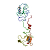
| ||||||||||
|---|---|---|---|---|---|---|---|---|---|---|---|
| 1 |
| ||||||||||
| Unit cell |
|
-Components
| #1: Protein | Mass: 20061.537 Da / Num. of mol.: 1 Source method: isolated from a genetically manipulated source Source: (gene. exp.) Homo sapiens (human) / Gene: HGF, HPTA / Production host:  | ||||||
|---|---|---|---|---|---|---|---|
| #2: Chemical |   Mass: 238.305 Da / Num. of mol.: 2 / Source method: obtained synthetically / Formula: C8H18N2O4S / Comment: pH buffer*YM Mass: 238.305 Da / Num. of mol.: 2 / Source method: obtained synthetically / Formula: C8H18N2O4S / Comment: pH buffer*YM#3: Water | ChemComp-HOH / |  Mass: 18.015 Da / Num. of mol.: 122 / Source method: isolated from a natural source / Formula: H2O Mass: 18.015 Da / Num. of mol.: 122 / Source method: isolated from a natural source / Formula: H2OHas ligand of interest | N | Has protein modification | Y | |
-Experimental details
-Experiment
| Experiment | Method: X-RAY DIFFRACTION / Number of used crystals: 1 |
|---|
- Sample preparation
| Crystal | Density Matthews: 2.2 Å3/Da / Density % sol: 44.16 % / Description: Thin hexagonal shaped |
|---|---|
| Crystal grow | Temperature: 290.15 K / Method: vapor diffusion, hanging drop / pH: 8.5 Details: 100 mM MOPS/HEPES pH 7.5, 30 mM sodium nitrate, 30 mM sodium phosphate dibasic, 30 mM ammonium sulphate, 20% v/v glycerol, 10% w/v PEG4000 Temp details: Inside a fridge-like incubator |
-Data collection
| Diffraction | Mean temperature: 100 K / Serial crystal experiment: N | ||||||||||||||||||||||||||||||
|---|---|---|---|---|---|---|---|---|---|---|---|---|---|---|---|---|---|---|---|---|---|---|---|---|---|---|---|---|---|---|---|
| Diffraction source | Source: SYNCHROTRON / Site: ESRF  / Beamline: ID23-1 / Wavelength: 0.9763 Å / Beamline: ID23-1 / Wavelength: 0.9763 Å | ||||||||||||||||||||||||||||||
| Detector | Type: DECTRIS PILATUS 6M-F / Detector: PIXEL / Date: Sep 29, 2014 / Details: Toroidal mirror | ||||||||||||||||||||||||||||||
| Radiation | Protocol: SINGLE WAVELENGTH / Monochromatic (M) / Laue (L): M / Scattering type: x-ray | ||||||||||||||||||||||||||||||
| Radiation wavelength | Wavelength: 0.9763 Å / Relative weight: 1 | ||||||||||||||||||||||||||||||
| Reflection | Resolution: 1.26→44.94 Å / Num. obs: 45262 / % possible obs: 99.8 % / Redundancy: 3.7 % / CC1/2: 0.998 / Rmerge(I) obs: 0.069 / Rpim(I) all: 0.041 / Rrim(I) all: 0.081 / Net I/σ(I): 8.4 / Num. measured all: 167746 | ||||||||||||||||||||||||||||||
| Reflection shell | Diffraction-ID: 1
|
- Processing
| Software |
| ||||||||||||||||
|---|---|---|---|---|---|---|---|---|---|---|---|---|---|---|---|---|---|
| Refinement | Method to determine structure: MOLECULAR REPLACEMENT Starting model: 1BHT Resolution: 1.7→35.89 Å / Cross valid method: THROUGHOUT
| ||||||||||||||||
| Solvent computation | Shrinkage radii: 0.9 Å / VDW probe radii: 1.11 Å | ||||||||||||||||
| Displacement parameters | Biso max: 67.43 Å2 / Biso mean: 22.03 Å2 / Biso min: 7.92 Å2 | ||||||||||||||||
| Refinement step | Cycle: LAST / Resolution: 1.7→35.89 Å
| ||||||||||||||||
| LS refinement shell | Resolution: 1.7→1.79 Å
|
