 Movie
Movie Controller


+ Open data

- Basic information
| Entry | Database: PDB / ID: 7o9i | |||||||||
|---|---|---|---|---|---|---|---|---|---|---|
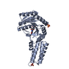
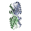
| Title | Escherichia coli Ffh in complex with pppGpp | |||||||||
 Components Components | Signal recognition particle protein | |||||||||
 Keywords Keywords | TRANSLATION / stringent response / targeting complex / signal recognition particle / alarmones / stress | |||||||||
| Function / homology |  Function and homology information Function and homology informationsignal recognition particle / signal-recognition-particle GTPase / 7S RNA binding / SRP-dependent cotranslational protein targeting to membrane / protein targeting to membrane / ribonucleoprotein complex / GTPase activity / GTP binding / cytosol Similarity search - Function | |||||||||
| Biological species |  | |||||||||
| Method |  X-RAY DIFFRACTION / SYNCHROTRON / MOLECULAR REPLACEMENT / Resolution: 2.49 Å X-RAY DIFFRACTION / SYNCHROTRON / MOLECULAR REPLACEMENT / Resolution: 2.49 Å | |||||||||
 Authors Authors | Czech, L. / Mais, C.-N. / Bange, G. | |||||||||
| Funding support |  Germany, 1items Germany, 1items
| |||||||||
 Citation Citation | Journal: Nat Commun / Year: 2022 Title: Inhibition of SRP-dependent protein secretion by the bacterial alarmone (p)ppGpp. Authors: Laura Czech / Christopher-Nils Mais / Hanna Kratzat / Pinku Sarmah / Pietro Giammarinaro / Sven-Andreas Freibert / Hanna Folke Esser / Joanna Musial / Otto Berninghausen / Wieland Steinchen ...Authors: Laura Czech / Christopher-Nils Mais / Hanna Kratzat / Pinku Sarmah / Pietro Giammarinaro / Sven-Andreas Freibert / Hanna Folke Esser / Joanna Musial / Otto Berninghausen / Wieland Steinchen / Roland Beckmann / Hans-Georg Koch / Gert Bange / Abstract: The stringent response enables bacteria to respond to nutrient limitation and other stress conditions through production of the nucleotide-based second messengers ppGpp and pppGpp, collectively known ...The stringent response enables bacteria to respond to nutrient limitation and other stress conditions through production of the nucleotide-based second messengers ppGpp and pppGpp, collectively known as (p)ppGpp. Here, we report that (p)ppGpp inhibits the signal recognition particle (SRP)-dependent protein targeting pathway, which is essential for membrane protein biogenesis and protein secretion. More specifically, (p)ppGpp binds to the SRP GTPases Ffh and FtsY, and inhibits the formation of the SRP receptor-targeting complex, which is central for the coordinated binding of the translating ribosome to the SecYEG translocon. Cryo-EM analysis of SRP bound to translating ribosomes suggests that (p)ppGpp may induce a distinct conformational stabilization of the NG domain of Ffh and FtsY in Bacillus subtilis but not in E. coli. | |||||||||
| History |
|
- Structure visualization
| Structure viewer | Molecule: MolmilJmol/JSmol |
|---|
- Downloads & links
-Download
| PDBx/mmCIF format | 7o9i.cif.gz | 70.8 KB | Display | PDBx/mmCIF format |
|---|---|---|---|---|
| PDB format | pdb7o9i.ent.gz | 49.8 KB | Display | PDB format |
| PDBx/mmJSON format | 7o9i.json.gz | Tree view | PDBx/mmJSON format | |
| Others |  Other downloads Other downloads |
-Validation report
| Arichive directory | https://data.pdbj.org/pub/pdb/validation_reports/o9/7o9iftp://data.pdbj.org/pub/pdb/validation_reports/o9/7o9i | HTTPS FTP |
|---|
-Related structure data
| Related structure data | 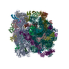 7o5bC  7o9fC  7o9gC  7o9hC 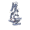 3ng1S S: Starting model for refinement C: citing same article ( |
|---|---|
| Similar structure data |


-Links
 PDBj
PDBj- Assembly
| Deposited unit | 
| ||||||||
|---|---|---|---|---|---|---|---|---|---|
| 1 |
| ||||||||
| Unit cell |
|
-Components
| #1: Protein | Mass: 33395.465 Da / Num. of mol.: 1 Source method: isolated from a genetically manipulated source Source: (gene. exp.) Strain: K12 / Gene: ffh, b2610, JW5414 / Production host: |
|---|---|
| #2: Chemical | ChemComp-0O2 /   Mass: 683.140 Da / Num. of mol.: 1 / Source method: obtained synthetically / Formula: C10H18N5O20P5 / Feature type: SUBJECT OF INVESTIGATION Mass: 683.140 Da / Num. of mol.: 1 / Source method: obtained synthetically / Formula: C10H18N5O20P5 / Feature type: SUBJECT OF INVESTIGATION |
| #3: Water | ChemComp-HOH /  Mass: 18.015 Da / Num. of mol.: 14 / Source method: isolated from a natural source / Formula: H2O Mass: 18.015 Da / Num. of mol.: 14 / Source method: isolated from a natural source / Formula: H2O |
| Has ligand of interest | Y |
-Experimental details
-Experiment
| Experiment | Method: X-RAY DIFFRACTION / Number of used crystals: 1 |
|---|
- Sample preparation
| Crystal | Density Matthews: 2.76 Å3/Da / Density % sol: 55.4 % |
|---|---|
| Crystal grow | Temperature: 293 K / Method: vapor diffusion, sitting drop / pH: 9.5 Details: 0.2 M sodium chloride, 0.1 M CHES pH 9.5, 50% PEG 400 |
-Data collection
| Diffraction | Mean temperature: 100 K / Serial crystal experiment: N |
|---|---|
| Diffraction source | Source: SYNCHROTRON / Site: ESRF  / Beamline: MASSIF-3 / Wavelength: 0.976253 Å / Beamline: MASSIF-3 / Wavelength: 0.976253 Å |
| Detector | Type: DECTRIS EIGER X 4M / Detector: PIXEL / Date: Sep 13, 2020 |
| Radiation | Protocol: SINGLE WAVELENGTH / Monochromatic (M) / Laue (L): M / Scattering type: x-ray |
| Radiation wavelength | Wavelength: 0.976253 Å / Relative weight: 1 |
| Reflection | Resolution: 2.49→41.05 Å / Num. obs: 11747 / % possible obs: 99.56 % / Redundancy: 19.2 % / CC1/2: 0.999 / Net I/σ(I): 25.77 |
| Reflection shell | Resolution: 2.49→2.58 Å / Redundancy: 14.4 % / Num. unique obs: 1112 / CC1/2: 0.969 / % possible all: 96.29 |
- Processing
| Software |
| |||||||||||||||||||||||||||||||||||
|---|---|---|---|---|---|---|---|---|---|---|---|---|---|---|---|---|---|---|---|---|---|---|---|---|---|---|---|---|---|---|---|---|---|---|---|---|
| Refinement | Method to determine structure: MOLECULAR REPLACEMENT Starting model: 3NG1 Resolution: 2.49→41.05 Å / SU ML: 0.35 / Cross valid method: FREE R-VALUE / σ(F): 1.38 / Phase error: 33.52 / Stereochemistry target values: ML
| |||||||||||||||||||||||||||||||||||
| Solvent computation | Shrinkage radii: 0.9 Å / VDW probe radii: 1.11 Å / Solvent model: FLAT BULK SOLVENT MODEL | |||||||||||||||||||||||||||||||||||
| Refinement step | Cycle: LAST / Resolution: 2.49→41.05 Å
| |||||||||||||||||||||||||||||||||||
| Refine LS restraints |
| |||||||||||||||||||||||||||||||||||
| LS refinement shell |
|
