 Movie
Movie Controller

[English] 日本語
 Yorodumi
- PDB-7o1b: Human phosphomannomutase 2 (PMM2) wild-type co-crystallized with ... -
+ Open data

- Basic information
| Entry | Database: PDB / ID: 7o1b | |||||||||
|---|---|---|---|---|---|---|---|---|---|---|
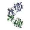
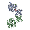
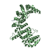
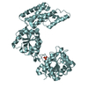
| Title | Human phosphomannomutase 2 (PMM2) wild-type co-crystallized with the activator glucose 1,6-bisphosphate | |||||||||
 Components Components | Phosphomannomutase 2 | |||||||||
 Keywords Keywords | ISOMERASE / glycobiology / congenital disorders of glycosylation / phosphotransferase / phosphomutase | |||||||||
| Function / homology |  Function and homology information Function and homology informationDefective PMM2 causes PMM2-CDG / : / : / Synthesis of GDP-mannose / phosphomannomutase / phosphomannomutase activity / GDP-mannose biosynthetic process / mannose metabolic process / glycoprotein biosynthetic process / protein N-linked glycosylation ...Defective PMM2 causes PMM2-CDG / : / : / Synthesis of GDP-mannose / phosphomannomutase / phosphomannomutase activity / GDP-mannose biosynthetic process / mannose metabolic process / glycoprotein biosynthetic process / protein N-linked glycosylation / ciliary tip / microtubule cytoskeleton / cilium / neuronal cell body / nucleoplasm / metal ion binding / cytosol Similarity search - Function | |||||||||
| Biological species |  Homo sapiens (human) Homo sapiens (human) | |||||||||
| Method |  X-RAY DIFFRACTION / SYNCHROTRON / MOLECULAR REPLACEMENT / Resolution: 3.08 Å X-RAY DIFFRACTION / SYNCHROTRON / MOLECULAR REPLACEMENT / Resolution: 3.08 Å | |||||||||
 Authors Authors | Ramon-Maiques, S. / Briso-Montiano, A. / Del Cano-Ochoa, F. / Vilas, A. / Perez, B. / Rubio, V. | |||||||||
| Funding support |  Spain, 2items Spain, 2items
| |||||||||
 Citation Citation | Journal: J Inherit Metab Dis / Year: 2022 Title: Insight on molecular pathogenesis and pharmacochaperoning potential in phosphomannomutase 2 deficiency, provided by novel human phosphomannomutase 2 structures. Authors: Briso-Montiano, A. / Del Cano-Ochoa, F. / Vilas, A. / Velazquez-Campoy, A. / Rubio, V. / Perez, B. / Ramon-Maiques, S. | |||||||||
| History |
|
- Structure visualization
| Structure viewer | Molecule: MolmilJmol/JSmol |
|---|
- Downloads & links
-Download
| PDBx/mmCIF format | 7o1b.cif.gz | 235 KB | Display | PDBx/mmCIF format |
|---|---|---|---|---|
| PDB format | pdb7o1b.ent.gz | 154.5 KB | Display | PDB format |
| PDBx/mmJSON format | 7o1b.json.gz | Tree view | PDBx/mmJSON format | |
| Others |  Other downloads Other downloads |
-Validation report
| Arichive directory | https://data.pdbj.org/pub/pdb/validation_reports/o1/7o1bftp://data.pdbj.org/pub/pdb/validation_reports/o1/7o1b | HTTPS FTP |
|---|
-Related structure data
| Related structure data |  7o0cSC  7o4gC 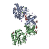 7o58C 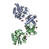 7o5zC S: Starting model for refinement C: citing same article ( |
|---|---|
| Similar structure data |


-Links
 PDBj
PDBj- Assembly
| Deposited unit | 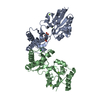
| |||||||||||||||
|---|---|---|---|---|---|---|---|---|---|---|---|---|---|---|---|---|
| 1 |
| |||||||||||||||
| Unit cell |
| |||||||||||||||
| Components on special symmetry positions |
|
-Components
| #1: Protein | Mass: 28274.336 Da / Num. of mol.: 2 Source method: isolated from a genetically manipulated source Details: Residues GPMAAP at the N-terminus are not seen in the electron density map. The most N-terminal GP sequence is part of the fusion tag after cleavage Source: (gene. exp.) Homo sapiens (human) / Gene: PMM2 / Plasmid: pOPIN-B / Production host:  #2: Chemical | ChemComp-MG /   Mass: 24.305 Da / Num. of mol.: 4 / Source method: obtained synthetically / Formula: Mg / Feature type: SUBJECT OF INVESTIGATION Mass: 24.305 Da / Num. of mol.: 4 / Source method: obtained synthetically / Formula: Mg / Feature type: SUBJECT OF INVESTIGATION#3: Sugar | ChemComp-G16 / |   Type: D-saccharide / Mass: 339.108 Da / Num. of mol.: 1 / Source method: obtained synthetically / Formula: C6H13O12P2 / Feature type: SUBJECT OF INVESTIGATION Type: D-saccharide / Mass: 339.108 Da / Num. of mol.: 1 / Source method: obtained synthetically / Formula: C6H13O12P2 / Feature type: SUBJECT OF INVESTIGATION#4: Chemical | ChemComp-CL / |   Mass: 35.453 Da / Num. of mol.: 1 / Source method: obtained synthetically / Formula: Cl / Feature type: SUBJECT OF INVESTIGATION Mass: 35.453 Da / Num. of mol.: 1 / Source method: obtained synthetically / Formula: Cl / Feature type: SUBJECT OF INVESTIGATION#5: Water | ChemComp-HOH / |  Mass: 18.015 Da / Num. of mol.: 55 / Source method: isolated from a natural source / Formula: H2O Mass: 18.015 Da / Num. of mol.: 55 / Source method: isolated from a natural source / Formula: H2OHas ligand of interest | Y | Has protein modification | Y | |
|---|
-Experimental details
-Experiment
| Experiment | Method: X-RAY DIFFRACTION / Number of used crystals: 1 |
|---|
- Sample preparation
| Crystal | Density Matthews: 2.3 Å3/Da / Density % sol: 46.5 % Description: Crystals of hexagonal morphology and 0.025 mm in the largest dimension |
|---|---|
| Crystal grow | Temperature: 293 K / Method: vapor diffusion, hanging drop / pH: 7.5 Details: Drops made by mixing 1 microliter of protein solution and 1 microliter of crystallization deposit solution. Protein solution contained 5 mg/ml protein and 3 mM glucose 1,6-bisphophate in 20 ...Details: Drops made by mixing 1 microliter of protein solution and 1 microliter of crystallization deposit solution. Protein solution contained 5 mg/ml protein and 3 mM glucose 1,6-bisphophate in 20 mM Hepes pH 7.5 and 0.2 M NaCl. Crystallization solution contained 0.3-0.4 M MgCl2, 24% PEG3350 and 0.1 M HEPES pH 7.5 |
-Data collection
| Diffraction | Mean temperature: 100 K / Serial crystal experiment: N |
|---|---|
| Diffraction source | Source: SYNCHROTRON / Site: ESRF  / Beamline: MASSIF-1 / Wavelength: 0.966 Å / Beamline: MASSIF-1 / Wavelength: 0.966 Å |
| Detector | Type: DECTRIS PILATUS 2M / Detector: PIXEL / Date: Mar 11, 2018 |
| Radiation | Protocol: SINGLE WAVELENGTH / Monochromatic (M) / Laue (L): M / Scattering type: x-ray |
| Radiation wavelength | Wavelength: 0.966 Å / Relative weight: 1 |
| Reflection | Resolution: 3.08→61.45 Å / Num. obs: 10988 / % possible obs: 99.9 % / Redundancy: 18.5 % / Biso Wilson estimate: 54 Å2 / CC1/2: 0.997 / Rmerge(I) obs: 0.341 / Rpim(I) all: 0.081 / Rrim(I) all: 0.35 / Net I/σ(I): 8.4 |
| Reflection shell | Resolution: 3.08→3.129 Å / Redundancy: 18.6 % / Rmerge(I) obs: 1.539 / Mean I/σ(I) obs: 2.2 / Num. unique obs: 514 / CC1/2: 0.784 / Rpim(I) all: 0.364 / % possible all: 100 |
- Processing
| Software |
| |||||||||||||||||||||||||||||||||||
|---|---|---|---|---|---|---|---|---|---|---|---|---|---|---|---|---|---|---|---|---|---|---|---|---|---|---|---|---|---|---|---|---|---|---|---|---|
| Refinement | Method to determine structure: MOLECULAR REPLACEMENT Starting model: 7O0C Resolution: 3.08→61.45 Å / SU ML: 0.3675 / Cross valid method: FREE R-VALUE / σ(F): 1.34 / Phase error: 23.2433 Stereochemistry target values: GeoStd + Monomer Library + CDL v1.2
| |||||||||||||||||||||||||||||||||||
| Solvent computation | Shrinkage radii: 0.9 Å / VDW probe radii: 1.11 Å / Solvent model: FLAT BULK SOLVENT MODEL | |||||||||||||||||||||||||||||||||||
| Displacement parameters | Biso mean: 43.24 Å2 | |||||||||||||||||||||||||||||||||||
| Refinement step | Cycle: LAST / Resolution: 3.08→61.45 Å
| |||||||||||||||||||||||||||||||||||
| Refine LS restraints |
| |||||||||||||||||||||||||||||||||||
| LS refinement shell |
|
