 Movie
Movie Controller

[English] 日本語
 Yorodumi
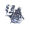
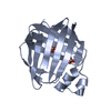
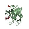
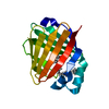
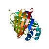
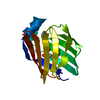
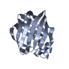
- PDB-7o0j: High resolution structure of recombinant chichen liver Bile Acid ... -
+ Open data

- Basic information
| Entry | Database: PDB / ID: 7o0j | ||||||
|---|---|---|---|---|---|---|---|
| Title | High resolution structure of recombinant chichen liver Bile Acid Binding Protein (cL-BABP) | ||||||
 Components Components | Fatty acid-binding protein, liver | ||||||
 Keywords Keywords | LIPID BINDING PROTEIN / fatty acid binding protein / chicken liver bile acid binding protein / recombinant | ||||||
| Function / homology |  Function and homology information Function and homology information | ||||||
| Biological species |  | ||||||
| Method |  X-RAY DIFFRACTION / SYNCHROTRON / MOLECULAR REPLACEMENT / Resolution: 1.4 Å X-RAY DIFFRACTION / SYNCHROTRON / MOLECULAR REPLACEMENT / Resolution: 1.4 Å | ||||||
 Authors Authors | Tassone, G. / Pozzi, C. | ||||||
 Citation Citation | Journal: Biomolecules / Year: 2021 Title: Validation of Recombinant Chicken Liver Bile Acid Binding Protein as a Tool for Cholic Acid Hosting. Authors: Tassone, G. / Orlandini, M. / Olivucci, M. / Pozzi, C. | ||||||
| History |
|
- Structure visualization
| Structure viewer | Molecule: MolmilJmol/JSmol |
|---|
- Downloads & links
-Download
| PDBx/mmCIF format | 7o0j.cif.gz | 74 KB | Display | PDBx/mmCIF format |
|---|---|---|---|---|
| PDB format | pdb7o0j.ent.gz | 53.1 KB | Display | PDB format |
| PDBx/mmJSON format | 7o0j.json.gz | Tree view | PDBx/mmJSON format | |
| Others |  Other downloads Other downloads |
-Validation report
| Arichive directory | https://data.pdbj.org/pub/pdb/validation_reports/o0/7o0jftp://data.pdbj.org/pub/pdb/validation_reports/o0/7o0j | HTTPS FTP |
|---|
-Related structure data
| Related structure data |  7o0kC  1tvqS S: Starting model for refinement C: citing same article ( |
|---|---|
| Similar structure data |


-Links
 PDBj
PDBj

- Assembly
| Deposited unit | 
| ||||||||
|---|---|---|---|---|---|---|---|---|---|
| 1 |
| ||||||||
| Unit cell |
|
-Components
| #1: Protein | Mass: 14513.648 Da / Num. of mol.: 1 Source method: isolated from a genetically manipulated source Source: (gene. exp.)  |
|---|---|
| #2: Water | ChemComp-HOH /  Mass: 18.015 Da / Num. of mol.: 212 / Source method: isolated from a natural source / Formula: H2O Mass: 18.015 Da / Num. of mol.: 212 / Source method: isolated from a natural source / Formula: H2O |
-Experimental details
-Experiment
| Experiment | Method: X-RAY DIFFRACTION / Number of used crystals: 1 |
|---|
- Sample preparation
| Crystal | Density Matthews: 2.89 Å3/Da / Density % sol: 57.48 % |
|---|---|
| Crystal grow | Temperature: 281 K / Method: vapor diffusion, sitting drop / pH: 7.5 Details: 10 % vol/vol 2-propanol, 20 % PEG4000 and 100 mM Hepes, pH 7.5 |
-Data collection
| Diffraction | Mean temperature: 100 K / Serial crystal experiment: N |
|---|---|
| Diffraction source | Source: SYNCHROTRON / Site: Diamond  / Beamline: I04 / Wavelength: 0.892 Å / Beamline: I04 / Wavelength: 0.892 Å |
| Detector | Type: DECTRIS EIGER2 X 16M / Detector: PIXEL / Date: Dec 4, 2019 |
| Radiation | Monochromator: Si(III) / Protocol: SINGLE WAVELENGTH / Monochromatic (M) / Laue (L): M / Scattering type: x-ray |
| Radiation wavelength | Wavelength: 0.892 Å / Relative weight: 1 |
| Reflection | Resolution: 1.4→71.29 Å / Num. obs: 32871 / % possible obs: 99.3 % / Redundancy: 9.3 % / CC1/2: 0.999 / Rmerge(I) obs: 0.038 / Rpim(I) all: 0.013 / Rrim(I) all: 0.04 / Net I/σ(I): 24.3 |
| Reflection shell | Resolution: 1.4→1.48 Å / Redundancy: 9.1 % / Rmerge(I) obs: 0.649 / Mean I/σ(I) obs: 3.1 / Num. unique obs: 4682 / CC1/2: 0.864 / Rpim(I) all: 0.225 / Rrim(I) all: 0.688 / % possible all: 98.5 |
- Processing
| Software |
| ||||||||||||||||||||||||||||||||||||||||||||||||||
|---|---|---|---|---|---|---|---|---|---|---|---|---|---|---|---|---|---|---|---|---|---|---|---|---|---|---|---|---|---|---|---|---|---|---|---|---|---|---|---|---|---|---|---|---|---|---|---|---|---|---|---|
| Refinement | Method to determine structure: MOLECULAR REPLACEMENT Starting model: 1TVQ Resolution: 1.4→32.55 Å / Cor.coef. Fo:Fc: 0.976 / Cor.coef. Fo:Fc free: 0.959 / SU B: 2.948 / SU ML: 0.052 / Cross valid method: THROUGHOUT / σ(F): 0 / ESU R: 0.056 / ESU R Free: 0.059 / Stereochemistry target values: MAXIMUM LIKELIHOOD / Details: U VALUES : REFINED INDIVIDUALLY
| ||||||||||||||||||||||||||||||||||||||||||||||||||
| Solvent computation | Ion probe radii: 0.8 Å / Shrinkage radii: 0.8 Å / VDW probe radii: 1.2 Å / Solvent model: MASK | ||||||||||||||||||||||||||||||||||||||||||||||||||
| Displacement parameters | Biso max: 292.04 Å2 / Biso mean: 30.033 Å2 / Biso min: 13.87 Å2
| ||||||||||||||||||||||||||||||||||||||||||||||||||
| Refine analyze | Luzzati coordinate error obs: 0.2001 Å | ||||||||||||||||||||||||||||||||||||||||||||||||||
| Refinement step | Cycle: final / Resolution: 1.4→32.55 Å
| ||||||||||||||||||||||||||||||||||||||||||||||||||
| Refine LS restraints |
| ||||||||||||||||||||||||||||||||||||||||||||||||||
| LS refinement shell | Resolution: 1.4→1.436 Å / Rfactor Rfree error: 0 / Total num. of bins used: 20
|
