 Movie
Movie Controller


+ Open data

- Basic information
| Entry | Database: PDB / ID: 7nzj | ||||||
|---|---|---|---|---|---|---|---|
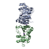
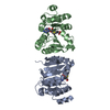
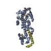
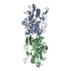
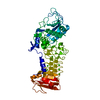
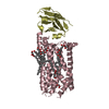
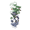
| Title | Structure of bsTrmB apo | ||||||
 Components Components | tRNA (guanine-N(7)-)-methyltransferase | ||||||
 Keywords Keywords | RNA BINDING PROTEIN / m7G Methyltransferase / Rossman-like fold / tRNA modifying enzyme | ||||||
| Function / homology |  Function and homology information Function and homology informationRNA (guanine-N7)-methylation / tRNA (guanine46-N7)-methyltransferase / tRNA (guanine(46)-N7)-methyltransferase activity / tRNA methyltransferase complex / tRNA methylation Similarity search - Function | ||||||
| Biological species |  | ||||||
| Method |  X-RAY DIFFRACTION / SYNCHROTRON / MOLECULAR REPLACEMENT / Resolution: 1.98 Å X-RAY DIFFRACTION / SYNCHROTRON / MOLECULAR REPLACEMENT / Resolution: 1.98 Å | ||||||
 Authors Authors | Blersch, K.F. / Ficner, R. / Neumann, P. | ||||||
| Funding support |  Germany, 1items Germany, 1items
| ||||||
 Citation Citation | Journal: Rna Biol. / Year: 2021 Title: Structural model of the M7G46 Methyltransferase TrmB in complex with tRNA. Authors: Blersch, K.F. / Burchert, J.P. / August, S.C. / Welp, L. / Neumann, P. / Koster, S. / Urlaub, H. / Ficner, R. | ||||||
| History |
|
- Structure visualization
| Structure viewer | Molecule: MolmilJmol/JSmol |
|---|
- Downloads & links
-Download
| PDBx/mmCIF format | 7nzj.cif.gz | 271.5 KB | Display | PDBx/mmCIF format |
|---|---|---|---|---|
| PDB format | pdb7nzj.ent.gz | 218.6 KB | Display | PDB format |
| PDBx/mmJSON format | 7nzj.json.gz | Tree view | PDBx/mmJSON format | |
| Others |  Other downloads Other downloads |
-Validation report
| Arichive directory | https://data.pdbj.org/pub/pdb/validation_reports/nz/7nzjftp://data.pdbj.org/pub/pdb/validation_reports/nz/7nzj | HTTPS FTP |
|---|
-Related structure data
| Related structure data |  7nybC 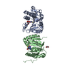 7nziC  2fcaS S: Starting model for refinement C: citing same article ( |
|---|---|
| Similar structure data |

-Links
 PDBj
PDBj

- Assembly
| Deposited unit | 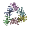
| ||||||||
|---|---|---|---|---|---|---|---|---|---|
| 1 | 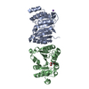
| ||||||||
| 2 | 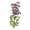
| ||||||||
| 3 | 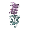
| ||||||||
| Unit cell |
|
-Components
| #1: Protein | Mass: 24537.744 Da / Num. of mol.: 6 Source method: isolated from a genetically manipulated source Source: (gene. exp.) Strain: 168 / Gene: trmB, ytmQ, BSU29900 / Production host: References: UniProt: O34522, tRNA (guanine46-N7)-methyltransferase #2: Chemical | ChemComp-NA / |   Mass: 22.990 Da / Num. of mol.: 1 / Source method: obtained synthetically / Formula: Na Mass: 22.990 Da / Num. of mol.: 1 / Source method: obtained synthetically / Formula: Na#3: Chemical | ChemComp-GOL /   Mass: 92.094 Da / Num. of mol.: 5 / Source method: obtained synthetically / Formula: C3H8O3 Mass: 92.094 Da / Num. of mol.: 5 / Source method: obtained synthetically / Formula: C3H8O3#4: Water | ChemComp-HOH / |  Mass: 18.015 Da / Num. of mol.: 912 / Source method: isolated from a natural source / Formula: H2O Mass: 18.015 Da / Num. of mol.: 912 / Source method: isolated from a natural source / Formula: H2OHas ligand of interest | N | |
|---|
-Experimental details
-Experiment
| Experiment | Method: X-RAY DIFFRACTION / Number of used crystals: 1 |
|---|
- Sample preparation
| Crystal | Density Matthews: 2.65 Å3/Da / Density % sol: 53.51 % |
|---|---|
| Crystal grow | Temperature: 293 K / Method: vapor diffusion, sitting drop / pH: 6 / Details: 0.2M NaCl, 0.1M MES, 15% v/v 5/4PO/OH, |
-Data collection
| Diffraction | Mean temperature: 100 K / Serial crystal experiment: N |
|---|---|
| Diffraction source | Source: SYNCHROTRON / Site: PETRA III, EMBL c/o DESY / Beamline: P13 (MX1) / Wavelength: 0.9763 Å |
| Detector | Type: DECTRIS PILATUS 6M-F / Detector: PIXEL / Date: Oct 27, 2019 |
| Radiation | Monochromator: M / Protocol: SINGLE WAVELENGTH / Monochromatic (M) / Laue (L): M / Scattering type: x-ray |
| Radiation wavelength | Wavelength: 0.9763 Å / Relative weight: 1 |
| Reflection | Resolution: 1.98→44.53 Å / Num. obs: 96395 / % possible obs: 91.5 % / Redundancy: 3.861 % / Biso Wilson estimate: 42.408 Å2 / CC1/2: 0.998 / Rmerge(I) obs: 0.072 / Rrim(I) all: 0.083 / Χ2: 0.85 / Net I/σ(I): 12.47 / Num. measured all: 372163 / Scaling rejects: 51 |
| Reflection shell | Resolution: 1.98→2 Å / Redundancy: 3.896 % / Rmerge(I) obs: 1.344 / Mean I/σ(I) obs: 1.08 / Num. unique obs: 2799 / CC1/2: 0.548 / Rrim(I) all: 1.557 / % possible all: 89.9 |
- Processing
| Software |
| ||||||||||||||||||||||||||||||||||||||||||||||||||||||||||||
|---|---|---|---|---|---|---|---|---|---|---|---|---|---|---|---|---|---|---|---|---|---|---|---|---|---|---|---|---|---|---|---|---|---|---|---|---|---|---|---|---|---|---|---|---|---|---|---|---|---|---|---|---|---|---|---|---|---|---|---|---|---|
| Refinement | Method to determine structure: MOLECULAR REPLACEMENT Starting model: 2FCA Resolution: 1.98→44.53 Å / Cor.coef. Fo:Fc: 0.966 / Cor.coef. Fo:Fc free: 0.951 / WRfactor Rfree: 0.2146 / WRfactor Rwork: 0.1808 / FOM work R set: 0.7547 / SU B: 5.5 / SU ML: 0.142 / SU R Cruickshank DPI: 0.1853 / SU Rfree: 0.1594 / Cross valid method: THROUGHOUT / σ(F): 0 / ESU R: 0.185 / ESU R Free: 0.159 / Stereochemistry target values: MAXIMUM LIKELIHOOD Details: HYDROGENS HAVE BEEN ADDED IN THE RIDING POSITIONS U VALUES : REFINED INDIVIDUALLY
| ||||||||||||||||||||||||||||||||||||||||||||||||||||||||||||
| Solvent computation | Ion probe radii: 0.8 Å / Shrinkage radii: 0.8 Å / VDW probe radii: 1.2 Å / Solvent model: MASK | ||||||||||||||||||||||||||||||||||||||||||||||||||||||||||||
| Displacement parameters | Biso max: 135.52 Å2 / Biso mean: 41.456 Å2 / Biso min: 25.9 Å2
| ||||||||||||||||||||||||||||||||||||||||||||||||||||||||||||
| Refinement step | Cycle: final / Resolution: 1.98→44.53 Å
| ||||||||||||||||||||||||||||||||||||||||||||||||||||||||||||
| Refine LS restraints |
| ||||||||||||||||||||||||||||||||||||||||||||||||||||||||||||
| LS refinement shell | Resolution: 1.98→2.031 Å / Rfactor Rfree error: 0 / Total num. of bins used: 20
|
