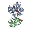
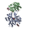
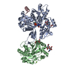
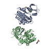
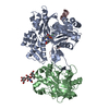
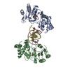
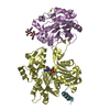
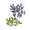
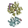
ジャーナル: Nat Struct Mol Biol / 年: 2021 タイトル: Higher-order phosphatase-substrate contacts terminate the integrated stress response. 著者: Yahui Yan / Heather P Harding / David Ron / 要旨: Many regulatory PPP1R subunits join few catalytic PP1c subunits to mediate phosphoserine and phosphothreonine dephosphorylation in metazoans. Regulatory subunits engage the surface of PP1c, locally ...Many regulatory PPP1R subunits join few catalytic PP1c subunits to mediate phosphoserine and phosphothreonine dephosphorylation in metazoans. Regulatory subunits engage the surface of PP1c, locally affecting flexible access of the phosphopeptide to the active site. However, catalytic efficiency of holophosphatases towards their phosphoprotein substrates remains unexplained. Here we present a cryo-EM structure of the tripartite PP1c-PPP1R15A-G-actin holophosphatase that terminates signaling in the mammalian integrated stress response (ISR) in the pre-dephosphorylation complex with its substrate, translation initiation factor 2α (eIF2α). G-actin, whose essential role in eIF2α dephosphorylation is supported crystallographically, biochemically and genetically, aligns the catalytic and regulatory subunits, creating a composite surface that engages the N-terminal domain of eIF2α to position the distant phosphoserine-51 at the active site. Substrate residues that mediate affinity for the holophosphatase also make critical contacts with eIF2α kinases. Thus, a convergent process of higher-order substrate recognition specifies functionally antagonistic phosphorylation and dephosphorylation in the ISR.
Proteinphosphatase1regulatorysubunit15A / Growth arrest and DNA damage-inducible protein GADD34 / Myeloid differentiation primary response ...Growth arrest and DNA damage-inducible protein GADD34 / Myeloid differentiation primary response protein MyD116 homolog
解像度: 2.55→55.11 Å / Cor.coef. Fo:Fc: 0.926 / Cor.coef. Fo:Fc free: 0.902 / SU B: 11.864 / SU ML: 0.241 / 交差検証法: THROUGHOUT / σ(F): 0 / ESU R: 0.491 / ESU R Free: 0.279 / 立体化学のターゲット値: MAXIMUM LIKELIHOOD 詳細: HYDROGENS HAVE BEEN ADDED IN THE RIDING POSITIONS U VALUES : REFINED INDIVIDUALLY
Rfactor
反射数
%反射
Selection details
Rfree
0.2478
2906
4.9 %
RANDOM
Rwork
0.2159
-
-
-
obs
0.2175
56449
99.85 %
-
溶媒の処理
イオンプローブ半径: 0.8 Å / 減衰半径: 0.8 Å / VDWプローブ半径: 1.2 Å / 溶媒モデル: MASK
 ムービー
ムービー コントローラー


 データを開く
 基本情報
基本情報 要素
要素 キーワード
キーワード 機能・相同性情報
 Homo sapiens (ヒト)
 X線回折 /
  データ登録者
 英国, 1件
  引用
引用 構造の表示
構造の表示 ダウンロードとリンク
 その他のダウンロード


 PDBj
PDBj


 集合体
集合体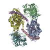


 分子量: 507.181 Da / 分子数: 2 / 由来タイプ: 合成 / 式: C10H16N5O13P3 / コメント: ATP, エネルギー貯蔵分子*YM
 分子量: 40.078 Da / 分子数: 6 / 由来タイプ: 合成 / 式: Ca
 試料調製
試料調製 解析
解析