 Movie
Movie Controller


+ Open data

- Basic information
| Entry | Database: PDB / ID: 7ntd | ||||||
|---|---|---|---|---|---|---|---|
| Title | The structure of the SBP TarP_Csal in complex with ferulate | ||||||
 Components Components | TRAP dicarboxylate transporter-DctP subunit | ||||||
 Keywords Keywords | TRANSPORT PROTEIN / TRAP transporter / solute binding protein / periplasmic / hydroxycinnamate / lignin | ||||||
| Function / homology | TRAP transporter solute receptor DctP / TRAP transporter solute receptor DctP superfamily / Bacterial extracellular solute-binding protein, family 7 / Twin arginine translocation (Tat) signal profile. / Twin-arginine translocation pathway, signal sequence / transmembrane transport / metal ion binding / 3-(4-HYDROXY-3-METHOXYPHENYL)-2-PROPENOIC ACID / TRAP dicarboxylate transporter-DctP subunit Function and homology information Function and homology information | ||||||
| Biological species |  Chromohalobacter salexigens (bacteria) Chromohalobacter salexigens (bacteria) | ||||||
| Method |  X-RAY DIFFRACTION / SYNCHROTRON / MOLECULAR REPLACEMENT / Resolution: 1.75 Å X-RAY DIFFRACTION / SYNCHROTRON / MOLECULAR REPLACEMENT / Resolution: 1.75 Å | ||||||
 Authors Authors | Bisson, C. / Salmon, R.C. / West, L. / Rafferty, J.B. / Hitchcock, A. / Thomas, G.H. / Kelly, D.J. | ||||||
| Funding support | 1items
| ||||||
 Citation Citation | Journal: Febs J. / Year: 2022 Title: The structural basis for high-affinity uptake of lignin-derived aromatic compounds by proteobacterial TRAP transporters. Authors: Bisson, C. / Salmon, R.C. / West, L. / Rafferty, J.B. / Hitchcock, A. / Thomas, G.H. / Kelly, D.J. | ||||||
| History |
|
- Structure visualization
| Structure viewer | Molecule: MolmilJmol/JSmol |
|---|
- Downloads & links
-Download
| PDBx/mmCIF format | 7ntd.cif.gz | 154.8 KB | Display | PDBx/mmCIF format |
|---|---|---|---|---|
| PDB format | pdb7ntd.ent.gz | Display | PDB format | |
| PDBx/mmJSON format | 7ntd.json.gz | Tree view | PDBx/mmJSON format | |
| Others |  Other downloads Other downloads |
-Validation report
| Arichive directory | https://data.pdbj.org/pub/pdb/validation_reports/nt/7ntdftp://data.pdbj.org/pub/pdb/validation_reports/nt/7ntd | HTTPS FTP |
|---|
-Related structure data
| Related structure data |  7nqgC  7nr2C  7nraC  7nrrC  7nswSC  7nteC C: citing same article ( S: Starting model for refinement |
|---|---|
| Similar structure data |


-Links
 PDBj
PDBj

- Assembly
| Deposited unit | 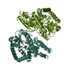
| ||||||||
|---|---|---|---|---|---|---|---|---|---|
| 1 | 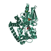
| ||||||||
| 2 | 
| ||||||||
| Unit cell |
|
-Components
| #1: Protein | Mass: 37303.809 Da / Num. of mol.: 2 Source method: isolated from a genetically manipulated source Details: C-terminal 6xHis tag and linker Source: (gene. exp.) Chromohalobacter salexigens (strain ATCC BAA-138 / DSM 3043 / CIP 106854 / NCIMB 13768 / 1H11) (bacteria)Strain: ATCC BAA-138 / DSM 3043 / CIP 106854 / NCIMB 13768 / 1H11 Gene: Csal_0280 / Production host: #2: Chemical |   Mass: 194.184 Da / Num. of mol.: 2 / Source method: obtained synthetically / Formula: C10H10O4 / Feature type: SUBJECT OF INVESTIGATION Mass: 194.184 Da / Num. of mol.: 2 / Source method: obtained synthetically / Formula: C10H10O4 / Feature type: SUBJECT OF INVESTIGATION#3: Chemical |   Mass: 24.305 Da / Num. of mol.: 2 / Source method: obtained synthetically / Formula: Mg Mass: 24.305 Da / Num. of mol.: 2 / Source method: obtained synthetically / Formula: Mg#4: Chemical | ChemComp-SO4 / |   Mass: 96.063 Da / Num. of mol.: 1 / Source method: obtained synthetically / Formula: SO4 Mass: 96.063 Da / Num. of mol.: 1 / Source method: obtained synthetically / Formula: SO4#5: Water | ChemComp-HOH / |  Mass: 18.015 Da / Num. of mol.: 472 / Source method: isolated from a natural source / Formula: H2O Mass: 18.015 Da / Num. of mol.: 472 / Source method: isolated from a natural source / Formula: H2OHas ligand of interest | Y | |
|---|
-Experimental details
-Experiment
| Experiment | Method: X-RAY DIFFRACTION / Number of used crystals: 1 |
|---|
- Sample preparation
| Crystal | Density Matthews: 2.21 Å3/Da / Density % sol: 44.44 % |
|---|---|
| Crystal grow | Temperature: 290 K / Method: vapor diffusion, hanging drop / pH: 8.5 Details: 0.2 M MgCl2, 0.1 M Tris pH 8.5 and 20 % (w/v) PEG 8000 |
-Data collection
| Diffraction | Mean temperature: 100 K / Serial crystal experiment: N |
|---|---|
| Diffraction source | Source: SYNCHROTRON / Site: Diamond  / Beamline: I04 / Wavelength: 0.97949 Å / Beamline: I04 / Wavelength: 0.97949 Å |
| Detector | Type: DECTRIS PILATUS 6M / Detector: PIXEL / Date: Jun 27, 2015 |
| Radiation | Protocol: SINGLE WAVELENGTH / Monochromatic (M) / Laue (L): M / Scattering type: x-ray |
| Radiation wavelength | Wavelength: 0.97949 Å / Relative weight: 1 |
| Reflection | Resolution: 1.75→48.89 Å / Num. obs: 62516 / % possible obs: 96.8 % / Redundancy: 2 % / CC1/2: 0.988 / Rmerge(I) obs: 0.08 / Rpim(I) all: 0.073 / Net I/σ(I): 6.2 |
| Reflection shell | Resolution: 1.75→1.8 Å / Redundancy: 2 % / Rmerge(I) obs: 0.687 / Num. unique obs: 4572 / CC1/2: 0.47 / Rpim(I) all: 0.62 / % possible all: 95.4 |
- Processing
| Software |
| ||||||||||||||||||||||||||||||||||||||||||||||||||||||||||||||||||||||||||||||||||||||||||||||||||||||||||||||||||||||||||||||||||||||||||||||||||||||||||||||||
|---|---|---|---|---|---|---|---|---|---|---|---|---|---|---|---|---|---|---|---|---|---|---|---|---|---|---|---|---|---|---|---|---|---|---|---|---|---|---|---|---|---|---|---|---|---|---|---|---|---|---|---|---|---|---|---|---|---|---|---|---|---|---|---|---|---|---|---|---|---|---|---|---|---|---|---|---|---|---|---|---|---|---|---|---|---|---|---|---|---|---|---|---|---|---|---|---|---|---|---|---|---|---|---|---|---|---|---|---|---|---|---|---|---|---|---|---|---|---|---|---|---|---|---|---|---|---|---|---|---|---|---|---|---|---|---|---|---|---|---|---|---|---|---|---|---|---|---|---|---|---|---|---|---|---|---|---|---|---|---|---|---|
| Refinement | Method to determine structure: MOLECULAR REPLACEMENT Starting model: 7NSW Resolution: 1.75→47.124 Å / Cor.coef. Fo:Fc: 0.961 / Cor.coef. Fo:Fc free: 0.937 / SU B: 3.876 / SU ML: 0.117 / Cross valid method: FREE R-VALUE / ESU R: 0.132 / ESU R Free: 0.138 Details: Hydrogens have been added in their riding positions
| ||||||||||||||||||||||||||||||||||||||||||||||||||||||||||||||||||||||||||||||||||||||||||||||||||||||||||||||||||||||||||||||||||||||||||||||||||||||||||||||||
| Solvent computation | Ion probe radii: 0.8 Å / Shrinkage radii: 0.8 Å / VDW probe radii: 1.2 Å / Solvent model: MASK BULK SOLVENT | ||||||||||||||||||||||||||||||||||||||||||||||||||||||||||||||||||||||||||||||||||||||||||||||||||||||||||||||||||||||||||||||||||||||||||||||||||||||||||||||||
| Displacement parameters | Biso mean: 33.081 Å2
| ||||||||||||||||||||||||||||||||||||||||||||||||||||||||||||||||||||||||||||||||||||||||||||||||||||||||||||||||||||||||||||||||||||||||||||||||||||||||||||||||
| Refinement step | Cycle: LAST / Resolution: 1.75→47.124 Å
| ||||||||||||||||||||||||||||||||||||||||||||||||||||||||||||||||||||||||||||||||||||||||||||||||||||||||||||||||||||||||||||||||||||||||||||||||||||||||||||||||
| Refine LS restraints |
| ||||||||||||||||||||||||||||||||||||||||||||||||||||||||||||||||||||||||||||||||||||||||||||||||||||||||||||||||||||||||||||||||||||||||||||||||||||||||||||||||
| LS refinement shell |
|
