 Movie
Movie Controller


+ Open data

- Basic information
| Entry | Database: PDB / ID: 7npu | |||||||||||||||
|---|---|---|---|---|---|---|---|---|---|---|---|---|---|---|---|---|
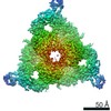
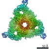
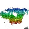
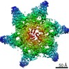
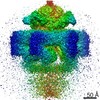
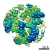
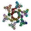
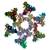
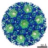
| Title | MycP5-free ESX-5 inner membrane complex, state I | |||||||||||||||
 Components Components |
| |||||||||||||||
 Keywords Keywords | MEMBRANE PROTEIN / T7SS / mycobacteria / protein transport / secretion / type VII secretion system / membrane | |||||||||||||||
| Function / homology |  Function and homology information Function and homology informationHydrolases; Acting on acid anhydrides / peptidoglycan-based cell wall / hydrolase activity / DNA binding / ATP binding / plasma membrane Similarity search - Function | |||||||||||||||
| Biological species |   Mycobacterium tuberculosis (bacteria) Mycobacterium tuberculosis (bacteria) | |||||||||||||||
| Method | ELECTRON MICROSCOPY / single particle reconstruction / cryo EM / Resolution: 4.48 Å | |||||||||||||||
 Authors Authors | Fahrenkamp, D. / Bunduc, C.M. / Wald, J. / Ummels, R. / Bitter, W. / Houben, E.N.G. / Marlovits, T.C. | |||||||||||||||
| Funding support |  Germany, Germany,  Netherlands, European Union, 4items Netherlands, European Union, 4items
| |||||||||||||||
 Citation Citation | Journal: Nature / Year: 2021 Title: Structure and dynamics of a mycobacterial type VII secretion system. Authors: Catalin M Bunduc / Dirk Fahrenkamp / Jiri Wald / Roy Ummels / Wilbert Bitter / Edith N G Houben / Thomas C Marlovits / Abstract: Mycobacterium tuberculosis is the cause of one of the most important infectious diseases in humans, which leads to 1.4 million deaths every year. Specialized protein transport systems-known as ...Mycobacterium tuberculosis is the cause of one of the most important infectious diseases in humans, which leads to 1.4 million deaths every year. Specialized protein transport systems-known as type VII secretion systems (T7SSs)-are central to the virulence of this pathogen, and are also crucial for nutrient and metabolite transport across the mycobacterial cell envelope. Here we present the structure of an intact T7SS inner-membrane complex of M. tuberculosis. We show how the 2.32-MDa ESX-5 assembly, which contains 165 transmembrane helices, is restructured and stabilized as a trimer of dimers by the MycP protease. A trimer of MycP caps a central periplasmic dome-like chamber that is formed by three EccB dimers, with the proteolytic sites of MycP facing towards the cavity. This chamber suggests a central secretion and processing conduit. Complexes without MycP show disruption of the EccB periplasmic assembly and increased flexibility, which highlights the importance of MycP for complex integrity. Beneath the EccB-MycP chamber, dimers of the EccC ATPase assemble into three bundles of four transmembrane helices each, which together seal the potential central secretion channel. Individual cytoplasmic EccC domains adopt two distinctive conformations that probably reflect different secretion states. Our work suggests a previously undescribed mechanism of protein transport and provides a structural scaffold to aid in the development of drugs against this major human pathogen. | |||||||||||||||
| History |
|
- Structure visualization
| Movie |
Movie viewer |
|---|---|
| Structure viewer | Molecule: MolmilJmol/JSmol |
- Downloads & links
-Download
| PDBx/mmCIF format | 7npu.cif.gz | 2.6 MB | Display | PDBx/mmCIF format |
|---|---|---|---|---|
| PDB format | pdb7npu.ent.gz | Display | PDB format | |
| PDBx/mmJSON format | 7npu.json.gz | Tree view | PDBx/mmJSON format | |
| Others |  Other downloads Other downloads |
-Validation report
| Arichive directory | https://data.pdbj.org/pub/pdb/validation_reports/np/7npuftp://data.pdbj.org/pub/pdb/validation_reports/np/7npu | HTTPS FTP |
|---|
-Related structure data
| Related structure data |  12521MC  7np7C  7nprC  7npsC  7nptC 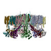 7npvC C: citing same article ( M: map data used to model this data |
|---|---|
| Similar structure data |


-Links
 PDBj
PDBj- Assembly
| Deposited unit | 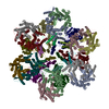
|
|---|---|
| 1 |
|
-Components
| #1: Protein | Mass: 53769.988 Da / Num. of mol.: 6 Source method: isolated from a genetically manipulated source Source: (gene. exp.) Mycobacterium tuberculosis (strain ATCC 25618 / H37Rv) (bacteria)Strain: ATCC 25618 / H37Rv / Gene: eccB5, Rv1782 Production host: Mycolicibacterium smegmatis MC2 155 (bacteria)References: UniProt: P9WNQ9, Hydrolases; Acting on acid anhydrides #2: Protein | Mass: 152900.750 Da / Num. of mol.: 6 Source method: isolated from a genetically manipulated source Source: (gene. exp.) Mycobacterium tuberculosis (strain ATCC 25618 / H37Rv) (bacteria)Strain: ATCC 25618 / H37Rv / Gene: eccC5, Rv1783 Production host: Mycolicibacterium smegmatis MC2 155 (bacteria)References: UniProt: P9WNA5 #3: Protein | Mass: 53480.906 Da / Num. of mol.: 12 Source method: isolated from a genetically manipulated source Source: (gene. exp.) Mycobacterium tuberculosis (strain ATCC 25618 / H37Rv) (bacteria)Strain: ATCC 25618 / H37Rv / Gene: eccD5, Rv1795 Production host: Mycolicibacterium smegmatis MC2 155 (bacteria)References: UniProt: P9WNP9 |
|---|
-Experimental details
-Experiment
| Experiment | Method: ELECTRON MICROSCOPY |
|---|---|
| EM experiment | Aggregation state: PARTICLE / 3D reconstruction method: single particle reconstruction |
- Sample preparation
| Component | Name: MycP5-free ESX-5 inner membrane complex, state I / Type: COMPLEX / Entity ID: all / Source: RECOMBINANT |
|---|---|
| Molecular weight | Experimental value: NO |
| Source (natural) | Organism: Mycobacterium tuberculosis H37Rv (bacteria) |
| Source (recombinant) | Organism: Mycolicibacterium smegmatis MC2 155 (bacteria) |
| Buffer solution | pH: 8 |
| Specimen | Embedding applied: NO / Shadowing applied: NO / Staining applied: NO / Vitrification applied: YES |
| Vitrification | Cryogen name: ETHANE-PROPANE |
- Electron microscopy imaging
| Experimental equipment |  Model: Titan Krios / Image courtesy: FEI Company |
|---|---|
| Microscopy | Model: TFS KRIOS |
| Electron gun | Electron source:  FIELD EMISSION GUN / Accelerating voltage: 300 kV / Illumination mode: SPOT SCAN FIELD EMISSION GUN / Accelerating voltage: 300 kV / Illumination mode: SPOT SCAN |
| Electron lens | Mode: BRIGHT FIELD |
| Image recording | Electron dose: 59.5 e/Å2 / Film or detector model: GATAN K3 BIOQUANTUM (6k x 4k) |
- Processing
| Software |
| ||||||||||||||||
|---|---|---|---|---|---|---|---|---|---|---|---|---|---|---|---|---|---|
| EM software |
| ||||||||||||||||
| CTF correction | Type: PHASE FLIPPING AND AMPLITUDE CORRECTION | ||||||||||||||||
| 3D reconstruction | Resolution: 4.48 Å / Resolution method: FSC 0.143 CUT-OFF / Num. of particles: 226793 / Symmetry type: POINT |
