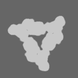
- EMDB-12519: Periplasmic assembly of the intact ESX-5 inner membrane complex, ... -
+
Open data
ID or keywords:
Loading...
-
Basic information
Entry
Database: EMDB / ID: EMD-12519
Title
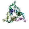
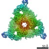
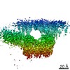
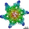
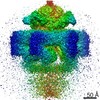
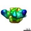
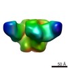
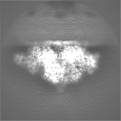
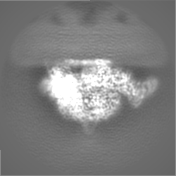
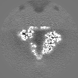
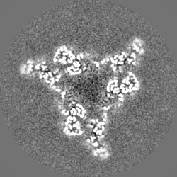
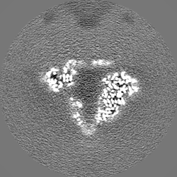
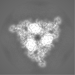
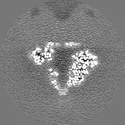
Periplasmic assembly of the intact ESX-5 inner membrane complex, C3 symmetry
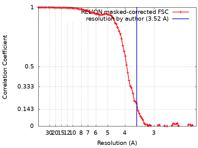
Map data
-50 Bfactor sharpening of 3D refinement map.
Sample
Complex: Periplasmic assembly of the intact ESX-5 inner membrane complex, C3 symmetry
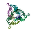
Protein or peptide: ESX-5 secretion system protein EccB5
Protein or peptide: ESX-5 secretion system protein MycP5
Function / homology
Function and homology information
Hydrolases; Acting on acid anhydrides / Hydrolases; Acting on peptide bonds (peptidases); Serine endopeptidases / peptidoglycan-based cell wall / protein processing / serine-type endopeptidase activity / hydrolase activity / ATP binding / plasma membrane Similarity search - Function
Type VII secretion system peptidase S8A, mycosin / Type VII secretion system EccB / Type VII secretion system EccB, repeat 3 domain / Type VII secretion system EccB, repeat 1 domain / Type VII secretion system ESX-1, transport TM domain B / Serine proteases, subtilase family, histidine active site. / Serine proteases, subtilase family, aspartic acid active site. / Peptidase S8, subtilisin, Asp-active site / Peptidase S8, subtilisin-related / Serine proteases, subtilase domain profile. ...Type VII secretion system peptidase S8A, mycosin / Type VII secretion system EccB / Type VII secretion system EccB, repeat 3 domain / Type VII secretion system EccB, repeat 1 domain / Type VII secretion system ESX-1, transport TM domain B / Serine proteases, subtilase family, histidine active site. / Serine proteases, subtilase family, aspartic acid active site. / Peptidase S8, subtilisin, Asp-active site / Peptidase S8, subtilisin-related / Serine proteases, subtilase domain profile. / Peptidase S8/S53 domain superfamily / Subtilase family / Peptidase S8/S53 domain Similarity search - Domain/homology
Netherlands Organisation for Scientific Research (NWO)
864.12.006
Netherlands
H2020 Marie Curie Actions of the European Commission
101030373
European Union
German Research Foundation (DFG)
FA1518/2-1
Germany
Citation
Journal: Nature / Year: 2021 Title: Structure and dynamics of a mycobacterial type VII secretion system. Authors: Catalin M Bunduc / Dirk Fahrenkamp / Jiri Wald / Roy Ummels / Wilbert Bitter / Edith N G Houben / Thomas C Marlovits / Abstract: Mycobacterium tuberculosis is the cause of one of the most important infectious diseases in humans, which leads to 1.4 million deaths every year. Specialized protein transport systems-known as ...Mycobacterium tuberculosis is the cause of one of the most important infectious diseases in humans, which leads to 1.4 million deaths every year. Specialized protein transport systems-known as type VII secretion systems (T7SSs)-are central to the virulence of this pathogen, and are also crucial for nutrient and metabolite transport across the mycobacterial cell envelope. Here we present the structure of an intact T7SS inner-membrane complex of M. tuberculosis. We show how the 2.32-MDa ESX-5 assembly, which contains 165 transmembrane helices, is restructured and stabilized as a trimer of dimers by the MycP protease. A trimer of MycP caps a central periplasmic dome-like chamber that is formed by three EccB dimers, with the proteolytic sites of MycP facing towards the cavity. This chamber suggests a central secretion and processing conduit. Complexes without MycP show disruption of the EccB periplasmic assembly and increased flexibility, which highlights the importance of MycP for complex integrity. Beneath the EccB-MycP chamber, dimers of the EccC ATPase assemble into three bundles of four transmembrane helices each, which together seal the potential central secretion channel. Individual cytoplasmic EccC domains adopt two distinctive conformations that probably reflect different secretion states. Our work suggests a previously undescribed mechanism of protein transport and provides a structural scaffold to aid in the development of drugs against this major human pathogen.
History
Deposition
Feb 28, 2021
-
Header (metadata) release
May 26, 2021
-
Map release
May 26, 2021
-
Update
Jun 2, 2021
-
Current status
Jun 2, 2021
Processing site: PDBe / Status: Released
-
Structure visualization
Movie
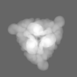
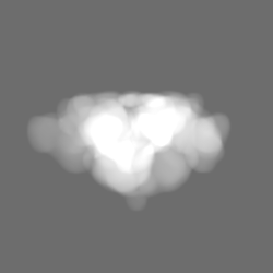
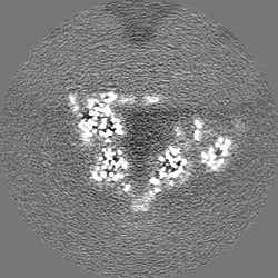
Surface view with section colored by density value
In the structure databanks used in Yorodumi, some data are registered as the other names, "COVID-19 virus" and "2019-nCoV". Here are the details of the virus and the list of structure data.
Jan 31, 2019. EMDB accession codes are about to change! (news from PDBe EMDB page)
EMDB accession codes are about to change! (news from PDBe EMDB page)
The allocation of 4 digits for EMDB accession codes will soon come to an end. Whilst these codes will remain in use, new EMDB accession codes will include an additional digit and will expand incrementally as the available range of codes is exhausted. The current 4-digit format prefixed with “EMD-” (i.e. EMD-XXXX) will advance to a 5-digit format (i.e. EMD-XXXXX), and so on. It is currently estimated that the 4-digit codes will be depleted around Spring 2019, at which point the 5-digit format will come into force.
The EM Navigator/Yorodumi systems omit the EMD- prefix.
Related info.:Q: What is EMD? / ID/Accession-code notation in Yorodumi/EM Navigator
Yorodumi is a browser for structure data from EMDB, PDB, SASBDB, etc.
This page is also the successor to EM Navigator detail page, and also detail information page/front-end page for Omokage search.
The word "yorodu" (or yorozu) is an old Japanese word meaning "ten thousand". "mi" (miru) is to see.
Related info.:EMDB / PDB / SASBDB / Comparison of 3 databanks / Yorodumi Search / Aug 31, 2016. New EM Navigator & Yorodumi / Yorodumi Papers / Jmol/JSmol / Function and homology information / Changes in new EM Navigator and Yorodumi
 Movie
Movie Controller

 Yorodumi
 Open data
 Basic information
 Map data
 Sample
 Function and homology information
 Mycobacterium tuberculosis H37Rv (bacteria)
 Authors
 Germany,
  Netherlands, European Union, 4 items
  Citation
 Structure visualization
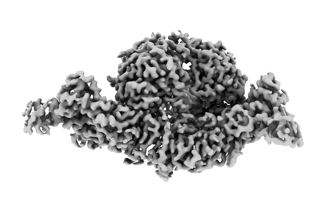
 Downloads & links
 emd_12519.png
 http://ftp.pdbj.org/pub/emdb/structures/EMD-12519


 Z (Sec.)
 Y (Row.)
 X (Col.)


 Sample components
 Processing
 Electron microscopy
 FIELD EMISSION GUN