 Movie
Movie Controller


+ Open data

- Basic information
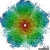
| Entry | Database: EMDB / ID: EMD-4016 | |||||||||
|---|---|---|---|---|---|---|---|---|---|---|
| Title | A 4.5 Angstrom structure of HIV-1 CA-SP1 | |||||||||
 Map data Map data | ||||||||||
 Sample Sample |
| |||||||||
| Biological species |   Human immunodeficiency virus 1 Human immunodeficiency virus 1 | |||||||||
| Method | subtomogram averaging / cryo EM / Resolution: 4.5 Å | |||||||||
 Authors Authors | Schur FKM / Obr M / Hagen WJH / Wan W / Arjen JJ / Kirkpatrick JM / Sachse C / Kraeusslich H-G / Briggs JAG | |||||||||
 Citation Citation | Journal: Science / Year: 2016 Title: An atomic model of HIV-1 capsid-SP1 reveals structures regulating assembly and maturation. Authors: Florian K M Schur / Martin Obr / Wim J H Hagen / William Wan / Arjen J Jakobi / Joanna M Kirkpatrick / Carsten Sachse / Hans-Georg Kräusslich / John A G Briggs /  Abstract: Immature HIV-1 assembles at and buds from the plasma membrane before proteolytic cleavage of the viral Gag polyprotein induces structural maturation. Maturation can be blocked by maturation ...Immature HIV-1 assembles at and buds from the plasma membrane before proteolytic cleavage of the viral Gag polyprotein induces structural maturation. Maturation can be blocked by maturation inhibitors (MIs), thereby abolishing infectivity. The CA (capsid) and SP1 (spacer peptide 1) region of Gag is the key regulator of assembly and maturation and is the target of MIs. We applied optimized cryo-electron tomography and subtomogram averaging to resolve this region within assembled immature HIV-1 particles at 3.9 angstrom resolution and built an atomic model. The structure reveals a network of intra- and intermolecular interactions mediating immature HIV-1 assembly. The proteolytic cleavage site between CA and SP1 is inaccessible to protease. We suggest that MIs prevent CA-SP1 cleavage by stabilizing the structure, and MI resistance develops by destabilizing CA-SP1. | |||||||||
| History |
|
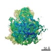
- Structure visualization
| Movie |
 Movie viewer Movie viewer |
|---|---|
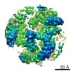
| Structure viewer | EM map: SurfViewMolmilJmol/JSmol |
| Supplemental images |

- Downloads & links
-EMDB archive
| Map data | emd_4016.map.gz | 24.7 MB | EMDB map data format | |
|---|---|---|---|---|
| Header (meta data) | emd-4016-v30.xmlemd-4016.xml | 15.2 KB 15.2 KB | Display Display | EMDB header |
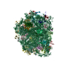
| Images | 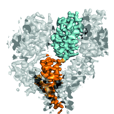 emd_4016.png emd_4016.png | 675.8 KB | ||
| Archive directory |  http://ftp.pdbj.org/pub/emdb/structures/EMD-4016ftp://ftp.pdbj.org/pub/emdb/structures/EMD-4016 http://ftp.pdbj.org/pub/emdb/structures/EMD-4016ftp://ftp.pdbj.org/pub/emdb/structures/EMD-4016 | HTTPS FTP |
-Related structure data
| Related structure data |  4015C  4017C  4018C 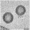 4019C 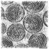 4020C 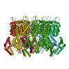 5l93C C: citing same article ( |
|---|---|
| Similar structure data |
-Links
| EMDB pages | EMDB (EBI/PDBe) / EMDataResource |
|---|---|
| Related items in Molecule of the Month |

-Map
| File | Download / File: emd_4016.map.gz / Format: CCP4 / Size: 27 MB / Type: IMAGE STORED AS FLOATING POINT NUMBER (4 BYTES) | ||||||||||||||||||||||||||||||||||||||||||||||||||||||||||||
|---|---|---|---|---|---|---|---|---|---|---|---|---|---|---|---|---|---|---|---|---|---|---|---|---|---|---|---|---|---|---|---|---|---|---|---|---|---|---|---|---|---|---|---|---|---|---|---|---|---|---|---|---|---|---|---|---|---|---|---|---|---|
| Projections & slices | Image control
Images are generated by Spider. | ||||||||||||||||||||||||||||||||||||||||||||||||||||||||||||
| Voxel size | X=Y=Z: 1.35 Å | ||||||||||||||||||||||||||||||||||||||||||||||||||||||||||||
| Density |
| ||||||||||||||||||||||||||||||||||||||||||||||||||||||||||||
| Symmetry | Space group: 1 | ||||||||||||||||||||||||||||||||||||||||||||||||||||||||||||
| Details | EMDB XML:
CCP4 map header:
| ||||||||||||||||||||||||||||||||||||||||||||||||||||||||||||
 Z (Sec.)
 Y (Row.)
 X (Col.)


-Supplemental data
- Sample components
-Entire : Human immunodeficiency virus 1
| Entire | Name: Human immunodeficiency virus 1 |
|---|---|
| Components |
|
-Supramolecule #1: Human immunodeficiency virus 1
| Supramolecule | Name: Human immunodeficiency virus 1 / type: virus / ID: 1 / Parent: 0 / Macromolecule list: all Details: Virus-like particles were obtained by in vitro assembly of a truncated Gag construct (deltaMACANCSP2) NCBI-ID: 11676 / Sci species name: Human immunodeficiency virus 1 / Virus type: VIRUS-LIKE PARTICLE / Virus isolate: OTHER / Virus enveloped: No / Virus empty: Yes |
|---|---|
| Host (natural) | Organism:  Homo sapiens (human) Homo sapiens (human) |
| Host system | Organism:  |
-Macromolecule #1: HIV-1 deltaMACANCSP2
| Macromolecule | Name: HIV-1 deltaMACANCSP2 / type: protein_or_peptide / ID: 1 / Enantiomer: LEVO |
|---|---|
| Sequence | String: SPRTLNAWVK VVEEKAFSPE VIPMFSALSE GATPQDLNTM LNTVGGHQAA MQMLKETINE EAAEWDRLH PVHAGPIAPG QMREPRGSDI AGTTSTLQEQ IGWMTHNPPI PVGEIYKRWI I LGLNKIVR MYSPTSILDI RQGPKEPFRD YVDRFYKTLR AEQASQEVKN ...String: SPRTLNAWVK VVEEKAFSPE VIPMFSALSE GATPQDLNTM LNTVGGHQAA MQMLKETINE EAAEWDRLH PVHAGPIAPG QMREPRGSDI AGTTSTLQEQ IGWMTHNPPI PVGEIYKRWI I LGLNKIVR MYSPTSILDI RQGPKEPFRD YVDRFYKTLR AEQASQEVKN WMTETLLVQN AN PDCKTIL KALGPGATLE EMMTACQGVG GPGHKARVLA EAMSQVT |
-Experimental details
-Structure determination
| Method | cryo EM |
|---|---|
 Processing Processing | subtomogram averaging |
| Aggregation state | particle |
-Sample preparation
| Concentration | 5 mg/mL | |||||||||||||||
|---|---|---|---|---|---|---|---|---|---|---|---|---|---|---|---|---|
| Buffer | pH: 8 Component:
Details: Virus-like particles were assembled in the presence of nucleic acid (73mer oligonucleotide, 1:10 molar ratio oligonucleotide:protein). | |||||||||||||||
| Grid | Model: C-flat / Material: COPPER / Mesh: 300 / Support film - Material: CARBON / Support film - topology: HOLEY / Pretreatment - Type: GLOW DISCHARGE / Details: at 20 mA | |||||||||||||||
| Vitrification | Cryogen name: ETHANE / Chamber humidity: 95 % / Chamber temperature: 15 K / Instrument: FEI VITROBOT MARK II Details: 10nM colloidal gold was added to the sample prior to plunge freezing.. | |||||||||||||||
| Details | Virus-like particles were assembled in vitro |
- Electron microscopy
| Microscope | FEI TITAN KRIOS |
|---|---|
| Specialist optics | Energy filter - Name: GIF Quantum LS / Energy filter - Lower energy threshold: 0 eV / Energy filter - Upper energy threshold: 20 eV |
| Details | Nanoprobe |
| Image recording | Film or detector model: GATAN K2 QUANTUM (4k x 4k) / Detector mode: SUPER-RESOLUTION / Digitization - Dimensions - Width: 3710 pixel / Digitization - Dimensions - Height: 3838 pixel / Digitization - Frames/image: 6-10 / Number grids imaged: 1 / Average exposure time: 1.0 sec. / Average electron dose: 5.0 e/Å2 Details: Number of frames ranged from 6-10 Exposure time per tilt ranged from 0.6 to 1.0 seconds |
| Electron beam | Acceleration voltage: 300 kV / Electron source:  FIELD EMISSION GUN FIELD EMISSION GUN |
| Electron optics | C2 aperture diameter: 50.0 µm / Illumination mode: FLOOD BEAM / Imaging mode: BRIGHT FIELD / Cs: 2.7 mm / Nominal defocus max: 4.5 µm / Nominal defocus min: 1.5 µm / Nominal magnification: 105000 |
| Sample stage | Specimen holder model: FEI TITAN KRIOS AUTOGRID HOLDER / Cooling holder cryogen: NITROGEN |
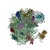
| Experimental equipment |  Model: Titan Krios / Image courtesy: FEI Company |
