 Movie
Movie Controller


+ Open data

- Basic information
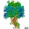
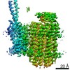
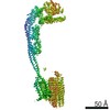
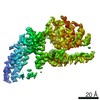
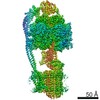
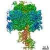
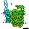
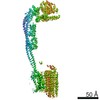
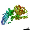
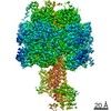
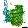
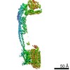
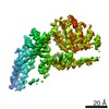
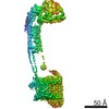
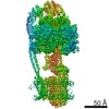
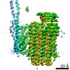
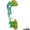
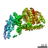
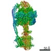
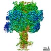
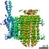
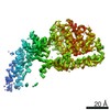
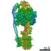
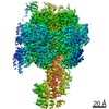
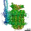
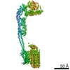
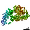
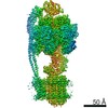
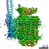
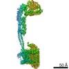
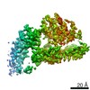
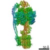
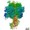
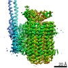
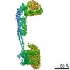
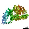
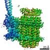
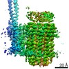
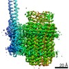
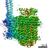
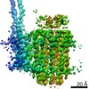
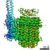
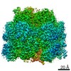
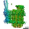
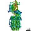
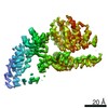
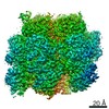
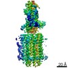
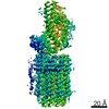
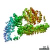
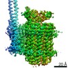
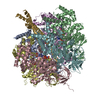
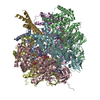
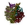
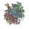
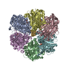
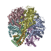
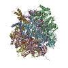
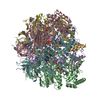
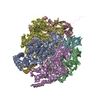
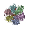
| Entry | Database: PDB / ID: 7nkh | |||||||||
|---|---|---|---|---|---|---|---|---|---|---|
| Title | Mycobacterium smegmatis ATP synthase F1 state 2 | |||||||||
 Components Components |
| |||||||||
 Keywords Keywords | HYDROLASE / complex / synthase | |||||||||
| Function / homology |  Function and homology information Function and homology informationproton motive force-driven plasma membrane ATP synthesis / H+-transporting two-sector ATPase / proton-transporting ATP synthase complex / proton-transporting ATP synthase activity, rotational mechanism / ADP binding / ATP hydrolysis activity / ATP binding / plasma membrane Similarity search - Function | |||||||||
| Biological species |  Mycolicibacterium smegmatis MC2 155 (bacteria) Mycolicibacterium smegmatis MC2 155 (bacteria) | |||||||||
| Method | ELECTRON MICROSCOPY / single particle reconstruction / cryo EM / Resolution: 2.78 Å | |||||||||
 Authors Authors | Montgomery, M.G. / Petri, J. / Spikes, T.E. / Walker, J.E. | |||||||||
| Funding support |  United Kingdom, 2items United Kingdom, 2items
| |||||||||
 Citation Citation | Journal: Proc Natl Acad Sci U S A / Year: 2021 Title: Structure of the ATP synthase from provides targets for treating tuberculosis. Authors: Martin G Montgomery / Jessica Petri / Tobias E Spikes / John E Walker / Abstract: The structure has been determined by electron cryomicroscopy of the adenosine triphosphate (ATP) synthase from This analysis confirms features in a prior description of the structure of the enzyme, ...The structure has been determined by electron cryomicroscopy of the adenosine triphosphate (ATP) synthase from This analysis confirms features in a prior description of the structure of the enzyme, but it also describes other highly significant attributes not recognized before that are crucial for understanding the mechanism and regulation of the mycobacterial enzyme. First, we resolved not only the three main states in the catalytic cycle described before but also eight substates that portray structural and mechanistic changes occurring during a 360° catalytic cycle. Second, a mechanism of auto-inhibition of ATP hydrolysis involves not only the engagement of the C-terminal region of an α-subunit in a loop in the γ-subunit, as proposed before, but also a "fail-safe" mechanism involving the b'-subunit in the peripheral stalk that enhances engagement. A third unreported characteristic is that the fused bδ-subunit contains a duplicated domain in its N-terminal region where the two copies of the domain participate in similar modes of attachment of the two of three N-terminal regions of the α-subunits. The auto-inhibitory plus the associated "fail-safe" mechanisms and the modes of attachment of the α-subunits provide targets for development of innovative antitubercular drugs. The structure also provides support for an observation made in the bovine ATP synthase that the transmembrane proton-motive force that provides the energy to drive the rotary mechanism is delivered directly and tangentially to the rotor via a Grotthuss water chain in a polar L-shaped tunnel. | |||||||||
| History |
|
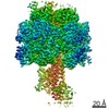
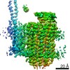
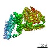
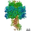
- Structure visualization
| Movie |
Movie viewer |
|---|---|
| Structure viewer | Molecule: MolmilJmol/JSmol |
- Downloads & links
-Download
| PDBx/mmCIF format | 7nkh.cif.gz | 922.4 KB | Display | PDBx/mmCIF format |
|---|---|---|---|---|
| PDB format | pdb7nkh.ent.gz | 778 KB | Display | PDB format |
| PDBx/mmJSON format | 7nkh.json.gz | Tree view | PDBx/mmJSON format | |
| Others |  Other downloads Other downloads |
-Validation report
| Summary document | 7nkh_validation.pdf.gz | 1.1 MB | Display | wwPDB validaton report |
|---|---|---|---|---|
| Full document | 7nkh_full_validation.pdf.gz | 1.1 MB | Display | |
| Data in XML | 7nkh_validation.xml.gz | 83.4 KB | Display | |
| Data in CIF | 7nkh_validation.cif.gz | 126.9 KB | Display | |
| Arichive directory | https://data.pdbj.org/pub/pdb/validation_reports/nk/7nkhftp://data.pdbj.org/pub/pdb/validation_reports/nk/7nkh | HTTPS FTP |
-Related structure data
| Related structure data | 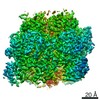 12439MC 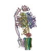 7njkC 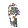 7njlC 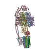 7njmC  7njnC 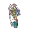 7njoC 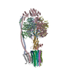 7njpC 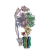 7njqC 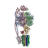 7njrC  7njsC 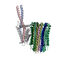 7njtC  7njuC  7njvC 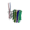 7njwC 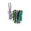 7njxC 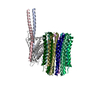 7njyC 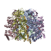 7nk7C 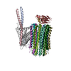 7nk9C 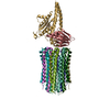 7nkbC 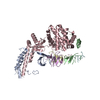 7nkdC 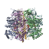 7nkjC  7nkkC 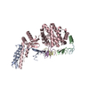 7nklC  7nknC 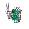 7nkpC 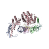 7nkqC 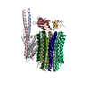 7nl9C M: map data used to model this data C: citing same article ( |
|---|---|
| Similar structure data |


-Links
 PDBj
PDBj


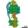
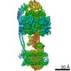
- Assembly
| Deposited unit | 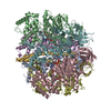
|
|---|---|
| 1 |
|
-Components
-ATP synthase subunit ... , 2 types, 6 molecules ABCDEF
| #1: Protein | Mass: 58951.461 Da / Num. of mol.: 3 Source method: isolated from a genetically manipulated source Source: (gene. exp.) Mycolicibacterium smegmatis MC2 155 (bacteria)Gene: atpA, MSMEG_4938, MSMEI_4811 / Production host: Mycolicibacterium smegmatis (bacteria) / Strain (production host): mc2 4517References: UniProt: A0R202, H+-transporting two-sector ATPase #2: Protein | Mass: 51670.453 Da / Num. of mol.: 3 Source method: isolated from a genetically manipulated source Source: (gene. exp.) Mycolicibacterium smegmatis MC2 155 (bacteria)Gene: atpD, MSMEG_4936, MSMEI_4809 / Production host: Mycolicibacterium smegmatis (bacteria) / Strain (production host): mc2 4517References: UniProt: A0R200, H+-transporting two-sector ATPase |
|---|
-Protein , 1 types, 1 molecules G
| #3: Protein | Mass: 33439.836 Da / Num. of mol.: 1 Source method: isolated from a genetically manipulated source Details: Alignment is incorrect. Source: (gene. exp.) Mycolicibacterium smegmatis MC2 155 (bacteria)Gene: atpG, MSMEG_4937, MSMEI_4810 / Production host: Mycolicibacterium smegmatis (bacteria) / Strain (production host): mc2 4517 / References: UniProt: A0R201 |
|---|
-Non-polymers , 4 types, 27 molecules 


| #4: Chemical | ChemComp-ATP /  Mass: 507.181 Da / Num. of mol.: 4 / Source method: obtained synthetically / Formula: C10H16N5O13P3 / Comment: ATP, energy-carrying molecule*YM Mass: 507.181 Da / Num. of mol.: 4 / Source method: obtained synthetically / Formula: C10H16N5O13P3 / Comment: ATP, energy-carrying molecule*YM#5: Chemical | ChemComp-MG /  Mass: 24.305 Da / Num. of mol.: 5 / Source method: obtained synthetically / Formula: Mg Mass: 24.305 Da / Num. of mol.: 5 / Source method: obtained synthetically / Formula: Mg#6: Chemical |  Mass: 427.201 Da / Num. of mol.: 2 / Source method: obtained synthetically / Formula: C10H15N5O10P2 / Comment: ADP, energy-carrying molecule*YM Mass: 427.201 Da / Num. of mol.: 2 / Source method: obtained synthetically / Formula: C10H15N5O10P2 / Comment: ADP, energy-carrying molecule*YM#7: Water | ChemComp-HOH / | Mass: 18.015 Da / Num. of mol.: 16 / Source method: isolated from a natural source / Formula: H2O |
|---|
-Details
| Has ligand of interest | N |
|---|
-Experimental details
-Experiment
| Experiment | Method: ELECTRON MICROSCOPY |
|---|---|
| EM experiment | Aggregation state: PARTICLE / 3D reconstruction method: single particle reconstruction |
- Sample preparation
| Component | Name: Mycobacterium smegmatis ATP synthase / Type: COMPLEX / Entity ID: #1-#3 / Source: RECOMBINANT |
|---|---|
| Molecular weight | Value: 0.547 MDa / Experimental value: YES |
| Source (natural) | Organism: Mycolicibacterium smegmatis MC2 155 (bacteria) |
| Source (recombinant) | Organism: Mycolicibacterium smegmatis (bacteria) / Strain: mc2 4517 |
| Buffer solution | pH: 8 |
| Specimen | Embedding applied: NO / Shadowing applied: NO / Staining applied: NO / Vitrification applied: YES |
| Vitrification | Cryogen name: ETHANE |
- Electron microscopy imaging
| Experimental equipment |  Model: Titan Krios / Image courtesy: FEI Company |
|---|---|
| Microscopy | Model: FEI TITAN KRIOS |
| Electron gun | Electron source:  FIELD EMISSION GUN / Accelerating voltage: 300 kV / Illumination mode: FLOOD BEAM FIELD EMISSION GUN / Accelerating voltage: 300 kV / Illumination mode: FLOOD BEAM |
| Electron lens | Mode: BRIGHT FIELD |
| Image recording | Electron dose: 59.86 e/Å2 / Film or detector model: GATAN K3 (6k x 4k) |
- Processing
| CTF correction | Type: PHASE FLIPPING AND AMPLITUDE CORRECTION |
|---|---|
| 3D reconstruction | Resolution: 2.78 Å / Resolution method: FSC 0.143 CUT-OFF / Num. of particles: 24938 / Symmetry type: POINT |
