 Movie
Movie Controller


+ Open data

- Basic information
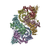
| Entry | Database: PDB / ID: 7ni6 | |||||||||||||||||||||||||||||||||||||||||||||||||||||||||||||||||||||||||||||||||||||||||||||
|---|---|---|---|---|---|---|---|---|---|---|---|---|---|---|---|---|---|---|---|---|---|---|---|---|---|---|---|---|---|---|---|---|---|---|---|---|---|---|---|---|---|---|---|---|---|---|---|---|---|---|---|---|---|---|---|---|---|---|---|---|---|---|---|---|---|---|---|---|---|---|---|---|---|---|---|---|---|---|---|---|---|---|---|---|---|---|---|---|---|---|---|---|---|---|
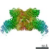
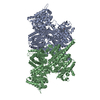
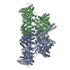
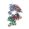
| Title | Human ATM kinase with bound ATPyS | |||||||||||||||||||||||||||||||||||||||||||||||||||||||||||||||||||||||||||||||||||||||||||||
 Components Components | Serine-protein kinase ATM | |||||||||||||||||||||||||||||||||||||||||||||||||||||||||||||||||||||||||||||||||||||||||||||
 Keywords Keywords | SIGNALING PROTEIN / Kinase / inhibitor / DNA damage response / cancer research | |||||||||||||||||||||||||||||||||||||||||||||||||||||||||||||||||||||||||||||||||||||||||||||
| Function / homology |  Function and homology information Function and homology informationestablishment of RNA localization to telomere / positive regulation of telomerase catalytic core complex assembly / cellular response to nitrosative stress / negative regulation of telomere capping / establishment of protein-containing complex localization to telomere / Sensing of DNA Double Strand Breaks / peptidyl-serine autophosphorylation / positive regulation of telomere maintenance via telomere lengthening / meiotic telomere clustering / DNA-dependent protein kinase activity ...establishment of RNA localization to telomere / positive regulation of telomerase catalytic core complex assembly / cellular response to nitrosative stress / negative regulation of telomere capping / establishment of protein-containing complex localization to telomere / Sensing of DNA Double Strand Breaks / peptidyl-serine autophosphorylation / positive regulation of telomere maintenance via telomere lengthening / meiotic telomere clustering / DNA-dependent protein kinase activity / extrinsic component of synaptic vesicle membrane / pre-B cell allelic exclusion / histone mRNA catabolic process / female meiotic nuclear division / regulation of telomere maintenance via telomerase / histone H2AXS139 kinase activity / male meiotic nuclear division / lipoprotein catabolic process / DNA double-strand break processing / V(D)J recombination / cellular response to X-ray / regulation of autophagosome assembly / pexophagy / Impaired BRCA2 binding to PALB2 / oocyte development / DNA repair complex / reciprocal meiotic recombination / positive regulation of DNA damage response, signal transduction by p53 class mediator / 1-phosphatidylinositol-3-kinase activity / TP53 Regulates Transcription of Caspase Activators and Caspases / HDR through Single Strand Annealing (SSA) / negative regulation of B cell proliferation / mitotic spindle assembly checkpoint signaling / cellular response to stress / response to ionizing radiation / positive regulation of double-strand break repair / Homologous DNA Pairing and Strand Exchange / Defective homologous recombination repair (HRR) due to BRCA1 loss of function / Defective HDR through Homologous Recombination Repair (HRR) due to PALB2 loss of BRCA1 binding function / Defective HDR through Homologous Recombination Repair (HRR) due to PALB2 loss of BRCA2/RAD51/RAD51C binding function / Resolution of D-loop Structures through Synthesis-Dependent Strand Annealing (SDSA) / Resolution of D-loop Structures through Holliday Junction Intermediates / TP53 Regulates Transcription of Genes Involved in Cytochrome C Release / mitotic G2 DNA damage checkpoint signaling / peroxisomal matrix / Impaired BRCA2 binding to RAD51 / replicative senescence / Regulation of HSF1-mediated heat shock response / Presynaptic phase of homologous DNA pairing and strand exchange / somitogenesis / ovarian follicle development / regulation of cellular response to heat / cellular response to retinoic acid / signal transduction in response to DNA damage / positive regulation of telomere maintenance via telomerase / negative regulation of TORC1 signaling / positive regulation of cell adhesion / DNA damage checkpoint signaling / telomere maintenance / thymus development / Pexophagy / regulation of signal transduction by p53 class mediator / determination of adult lifespan / regulation of autophagy / post-embryonic development / TP53 Regulates Transcription of DNA Repair Genes / DNA damage response, signal transduction by p53 class mediator / cellular response to reactive oxygen species / Nonhomologous End-Joining (NHEJ) / Stabilization of p53 / cellular response to gamma radiation / Autodegradation of the E3 ubiquitin ligase COP1 / brain development / G2/M DNA damage checkpoint / Regulation of TP53 Activity through Methylation / double-strand break repair via homologous recombination / DNA Damage/Telomere Stress Induced Senescence / double-strand break repair via nonhomologous end joining / Meiotic recombination / HDR through Homologous Recombination (HRR) / multicellular organism growth / spindle / intrinsic apoptotic signaling pathway in response to DNA damage / cellular senescence / Regulation of TP53 Degradation / protein autophosphorylation / positive regulation of neuron apoptotic process / double-strand break repair / chromosome / Recruitment and ATM-mediated phosphorylation of repair and signaling proteins at DNA double strand breaks / site of double-strand break / heart development / Processing of DNA double-strand break ends / neuron apoptotic process / regulation of apoptotic process / Regulation of TP53 Activity through Phosphorylation / protein phosphorylation / non-specific serine/threonine protein kinase / regulation of cell cycle / protein stabilization Similarity search - Function | |||||||||||||||||||||||||||||||||||||||||||||||||||||||||||||||||||||||||||||||||||||||||||||
| Biological species |  Homo sapiens (human) Homo sapiens (human) | |||||||||||||||||||||||||||||||||||||||||||||||||||||||||||||||||||||||||||||||||||||||||||||
| Method | ELECTRON MICROSCOPY / single particle reconstruction / cryo EM / Resolution: 2.8 Å | |||||||||||||||||||||||||||||||||||||||||||||||||||||||||||||||||||||||||||||||||||||||||||||
 Authors Authors | Stakyte, K. / Rotheneder, M. / Lammens, K. / Bartho, J.D. | |||||||||||||||||||||||||||||||||||||||||||||||||||||||||||||||||||||||||||||||||||||||||||||
| Funding support |  Germany, 4items Germany, 4items
| |||||||||||||||||||||||||||||||||||||||||||||||||||||||||||||||||||||||||||||||||||||||||||||
 Citation Citation | Journal: Nat Struct Mol Biol / Year: 2021 Title: Molecular basis of human ATM kinase inhibition. Authors: K Stakyte / M Rotheneder / K Lammens / J D Bartho / U Grädler / T Fuchß / U Pehl / A Alt / E van de Logt / K P Hopfner / Abstract: Human checkpoint kinase ataxia telangiectasia-mutated (ATM) plays a key role in initiation of the DNA damage response following DNA double-strand breaks. ATM inhibition is a promising approach in ...Human checkpoint kinase ataxia telangiectasia-mutated (ATM) plays a key role in initiation of the DNA damage response following DNA double-strand breaks. ATM inhibition is a promising approach in cancer therapy, but, so far, detailed insights into the binding modes of known ATM inhibitors have been hampered due to the lack of high-resolution ATM structures. Using cryo-EM, we have determined the structure of human ATM to an overall resolution sufficient to build a near-complete atomic model and identify two hitherto unknown zinc-binding motifs. We determined the structure of the kinase domain bound to ATPγS and to the ATM inhibitors KU-55933 and M4076 at 2.8 Å, 2.8 Å and 3.0 Å resolution, respectively. The mode of action and selectivity of the ATM inhibitors can be explained by structural comparison and provide a framework for structure-based drug design. | |||||||||||||||||||||||||||||||||||||||||||||||||||||||||||||||||||||||||||||||||||||||||||||
| History |
|
- Structure visualization
| Movie |
Movie viewer |
|---|---|
| Structure viewer | Molecule: MolmilJmol/JSmol |
- Downloads & links
-Download
| PDBx/mmCIF format | 7ni6.cif.gz | 575.5 KB | Display | PDBx/mmCIF format |
|---|---|---|---|---|
| PDB format | pdb7ni6.ent.gz | 427 KB | Display | PDB format |
| PDBx/mmJSON format | 7ni6.json.gz | Tree view | PDBx/mmJSON format | |
| Others |  Other downloads Other downloads |
-Validation report
| Arichive directory | https://data.pdbj.org/pub/pdb/validation_reports/ni/7ni6ftp://data.pdbj.org/pub/pdb/validation_reports/ni/7ni6 | HTTPS FTP |
|---|
-Related structure data
| Related structure data |  12352MC  7ni4C  7ni5C C: citing same article ( M: map data used to model this data |
|---|---|
| Similar structure data |


-Links
 PDBj
PDBj


- Assembly
| Deposited unit | 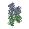
|
|---|---|
| 1 |
|
-Components
| #1: Protein | Mass: 354526.188 Da / Num. of mol.: 2 Source method: isolated from a genetically manipulated source Source: (gene. exp.) Homo sapiens (human) / Gene: ATM / Production host: Homo sapiens (human)References: UniProt: Q13315, non-specific serine/threonine protein kinase #2: Chemical |   Mass: 65.409 Da / Num. of mol.: 2 / Source method: obtained synthetically / Formula: Zn Mass: 65.409 Da / Num. of mol.: 2 / Source method: obtained synthetically / Formula: Zn#3: Chemical |   Mass: 523.247 Da / Num. of mol.: 2 / Source method: obtained synthetically / Formula: C10H16N5O12P3S / Feature type: SUBJECT OF INVESTIGATION / Comment: ATP-gamma-S, energy-carrying molecule analogue*YM Mass: 523.247 Da / Num. of mol.: 2 / Source method: obtained synthetically / Formula: C10H16N5O12P3S / Feature type: SUBJECT OF INVESTIGATION / Comment: ATP-gamma-S, energy-carrying molecule analogue*YM#4: Chemical |   Mass: 24.305 Da / Num. of mol.: 2 / Source method: obtained synthetically / Formula: Mg Mass: 24.305 Da / Num. of mol.: 2 / Source method: obtained synthetically / Formula: MgHas ligand of interest | Y | Has protein modification | N | |
|---|
-Experimental details
-Experiment
| Experiment | Method: ELECTRON MICROSCOPY |
|---|---|
| EM experiment | Aggregation state: PARTICLE / 3D reconstruction method: single particle reconstruction |
- Sample preparation
| Component | Name: ATM dimer with bound KU-55933 / Type: COMPLEX / Entity ID: #1 / Source: RECOMBINANT |
|---|---|
| Source (natural) | Organism: Homo sapiens (human) |
| Source (recombinant) | Organism: Homo sapiens (human) |
| Buffer solution | pH: 7.5 |
| Specimen | Embedding applied: NO / Shadowing applied: NO / Staining applied: NO / Vitrification applied: YES |
| Vitrification | Cryogen name: ETHANE |
- Electron microscopy imaging
| Experimental equipment |  Model: Titan Krios / Image courtesy: FEI Company |
|---|---|
| Microscopy | Model: FEI TITAN KRIOS |
| Electron gun | Electron source:  FIELD EMISSION GUN / Accelerating voltage: 300 kV / Illumination mode: FLOOD BEAM FIELD EMISSION GUN / Accelerating voltage: 300 kV / Illumination mode: FLOOD BEAM |
| Electron lens | Mode: BRIGHT FIELD |
| Image recording | Electron dose: 42 e/Å2 / Detector mode: COUNTING / Film or detector model: GATAN K2 SUMMIT (4k x 4k) |
- Processing
| Software | Name: PHENIX / Version: 1.19.1_4122: / Classification: refinement | ||||||||||||||||||||||||
|---|---|---|---|---|---|---|---|---|---|---|---|---|---|---|---|---|---|---|---|---|---|---|---|---|---|
| EM software |
| ||||||||||||||||||||||||
| CTF correction | Type: PHASE FLIPPING AND AMPLITUDE CORRECTION | ||||||||||||||||||||||||
| 3D reconstruction | Resolution: 2.8 Å / Resolution method: FSC 0.143 CUT-OFF / Num. of particles: 690548 / Symmetry type: POINT | ||||||||||||||||||||||||
| Refine LS restraints |
|
