 Movie
Movie Controller

[English] 日本語
 Yorodumi
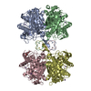
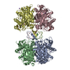
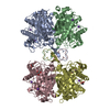
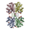
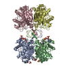
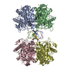
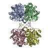
- PDB-7n7z: Structure of Acetyl-CoA acetyltransferase from Syntrophomonas wolfei -
+ Open data

- Basic information
| Entry | Database: PDB / ID: 7n7z | ||||||
|---|---|---|---|---|---|---|---|
| Title | Structure of Acetyl-CoA acetyltransferase from Syntrophomonas wolfei | ||||||
 Components Components | Acetyl-CoA C-acetyltransferase | ||||||
 Keywords Keywords | TRANSFERASE / Acetyl-CoA / Syntroph | ||||||
| Function / homology |  Function and homology information Function and homology informationacetyl-CoA C-acetyltransferase / acetyl-CoA C-acetyltransferase activity Similarity search - Function | ||||||
| Biological species |  Syntrophomonas wolfei subsp. wolfei (bacteria) Syntrophomonas wolfei subsp. wolfei (bacteria) | ||||||
| Method |  X-RAY DIFFRACTION / SYNCHROTRON / MOLECULAR REPLACEMENT / Resolution: 2.02 Å X-RAY DIFFRACTION / SYNCHROTRON / MOLECULAR REPLACEMENT / Resolution: 2.02 Å | ||||||
 Authors Authors | Arbing, M.A. / Gunsalus, R.P. | ||||||
| Funding support |  United States, 1items United States, 1items
| ||||||
 Citation Citation | Journal: Front Microbiol / Year: 2022 Title: Dynamic acylome reveals metabolite driven modifications in Syntrophomonas wolfei. Authors: Fu, J.Y. / Muroski, J.M. / Arbing, M.A. / Salguero, J.A. / Wofford, N.Q. / McInerney, M.J. / Gunsalus, R.P. / Loo, J.A. / Ogorzalek Loo, R.R. | ||||||
| History |
|
- Structure visualization
| Structure viewer | Molecule: MolmilJmol/JSmol |
|---|
- Downloads & links
-Download
| PDBx/mmCIF format | 7n7z.cif.gz | 515.3 KB | Display | PDBx/mmCIF format |
|---|---|---|---|---|
| PDB format | pdb7n7z.ent.gz | 359.6 KB | Display | PDB format |
| PDBx/mmJSON format | 7n7z.json.gz | Tree view | PDBx/mmJSON format | |
| Others |  Other downloads Other downloads |
-Validation report
| Arichive directory | https://data.pdbj.org/pub/pdb/validation_reports/n7/7n7zftp://data.pdbj.org/pub/pdb/validation_reports/n7/7n7z | HTTPS FTP |
|---|
-Related structure data
| Related structure data |  4wysS S: Starting model for refinement |
|---|---|
| Similar structure data |


-Links
 PDBj
PDBj

- Assembly
| Deposited unit | 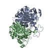
| ||||||||||||
|---|---|---|---|---|---|---|---|---|---|---|---|---|---|
| 1 | 
| ||||||||||||
| Unit cell |
|
-Components
| #1: Protein | Mass: 42268.012 Da / Num. of mol.: 2 Source method: isolated from a genetically manipulated source Source: (gene. exp.) Syntrophomonas wolfei subsp. wolfei (strain DSM 2245B / Goettingen) (bacteria)Strain: DSM 2245B / Goettingen / Gene: Swol_0675 / Plasmid: pMAPLe4 / Production host: #2: Water | ChemComp-HOH / |  Mass: 18.015 Da / Num. of mol.: 219 / Source method: isolated from a natural source / Formula: H2O Mass: 18.015 Da / Num. of mol.: 219 / Source method: isolated from a natural source / Formula: H2O |
|---|
-Experimental details
-Experiment
| Experiment | Method: X-RAY DIFFRACTION / Number of used crystals: 1 |
|---|
- Sample preparation
| Crystal | Density Matthews: 2.35 Å3/Da / Density % sol: 47.77 % |
|---|---|
| Crystal grow | Temperature: 293 K / Method: vapor diffusion, hanging drop / pH: 7 Details: Protein buffer: 20 mM Tris pH 8, 150 mM NaCl, 5 mM B-ME. Crystallization buffer: 0.1 M Sodium formate pH 7.0, 12% PEG 3350. Cryo buffer: 23% PEG 3350, 5% PEG 400. |
-Data collection
| Diffraction | Mean temperature: 100 K / Serial crystal experiment: N |
|---|---|
| Diffraction source | Source: SYNCHROTRON / Site: APS / Beamline: 24-ID-E / Wavelength: 0.97918 Å |
| Detector | Type: DECTRIS EIGER X 16M / Detector: PIXEL / Date: Oct 15, 2020 |
| Radiation | Protocol: SINGLE WAVELENGTH / Monochromatic (M) / Laue (L): M / Scattering type: x-ray |
| Radiation wavelength | Wavelength: 0.97918 Å / Relative weight: 1 |
| Reflection | Resolution: 2.02→41.64 Å / Num. obs: 53598 / % possible obs: 99.4 % / Redundancy: 5.9 % / Biso Wilson estimate: 33 Å2 / CC1/2: 0.997 / CC star: 0.999 / Rmerge(I) obs: 0.1041 / Rpim(I) all: 0.04584 / Rrim(I) all: 0.1141 / Net I/σ(I): 10.47 |
| Reflection shell | Resolution: 2.02→2.094 Å / Redundancy: 5.1 % / Rmerge(I) obs: 0.8022 / Mean I/σ(I) obs: 1.63 / Num. unique obs: 5118 / CC1/2: 0.511 / CC star: 0.822 / Rpim(I) all: 0.3867 / Rrim(I) all: 0.8933 / % possible all: 96.93 |
- Processing
| Software |
| |||||||||||||||||||||||||||||||||||||||||||||||||||||||||||||||||||||||||||||||||||||||||||||||||||||||||||||||||||||||||||||||||||||||||||||||||||||||||||||||||||||||||||||||||||||||||||||||||||||||||||||||||||||||||
|---|---|---|---|---|---|---|---|---|---|---|---|---|---|---|---|---|---|---|---|---|---|---|---|---|---|---|---|---|---|---|---|---|---|---|---|---|---|---|---|---|---|---|---|---|---|---|---|---|---|---|---|---|---|---|---|---|---|---|---|---|---|---|---|---|---|---|---|---|---|---|---|---|---|---|---|---|---|---|---|---|---|---|---|---|---|---|---|---|---|---|---|---|---|---|---|---|---|---|---|---|---|---|---|---|---|---|---|---|---|---|---|---|---|---|---|---|---|---|---|---|---|---|---|---|---|---|---|---|---|---|---|---|---|---|---|---|---|---|---|---|---|---|---|---|---|---|---|---|---|---|---|---|---|---|---|---|---|---|---|---|---|---|---|---|---|---|---|---|---|---|---|---|---|---|---|---|---|---|---|---|---|---|---|---|---|---|---|---|---|---|---|---|---|---|---|---|---|---|---|---|---|---|---|---|---|---|---|---|---|---|---|---|---|---|---|---|---|---|
| Refinement | Method to determine structure: MOLECULAR REPLACEMENT Starting model: 4WYS Resolution: 2.02→41.64 Å / SU ML: 0.3131 / Cross valid method: FREE R-VALUE / σ(F): 1.35 / Phase error: 28.765 Stereochemistry target values: GeoStd + Monomer Library + CDL v1.2
| |||||||||||||||||||||||||||||||||||||||||||||||||||||||||||||||||||||||||||||||||||||||||||||||||||||||||||||||||||||||||||||||||||||||||||||||||||||||||||||||||||||||||||||||||||||||||||||||||||||||||||||||||||||||||
| Solvent computation | Shrinkage radii: 0.9 Å / VDW probe radii: 1.11 Å / Solvent model: FLAT BULK SOLVENT MODEL | |||||||||||||||||||||||||||||||||||||||||||||||||||||||||||||||||||||||||||||||||||||||||||||||||||||||||||||||||||||||||||||||||||||||||||||||||||||||||||||||||||||||||||||||||||||||||||||||||||||||||||||||||||||||||
| Displacement parameters | Biso mean: 42.66 Å2 | |||||||||||||||||||||||||||||||||||||||||||||||||||||||||||||||||||||||||||||||||||||||||||||||||||||||||||||||||||||||||||||||||||||||||||||||||||||||||||||||||||||||||||||||||||||||||||||||||||||||||||||||||||||||||
| Refinement step | Cycle: LAST / Resolution: 2.02→41.64 Å
| |||||||||||||||||||||||||||||||||||||||||||||||||||||||||||||||||||||||||||||||||||||||||||||||||||||||||||||||||||||||||||||||||||||||||||||||||||||||||||||||||||||||||||||||||||||||||||||||||||||||||||||||||||||||||
| Refine LS restraints |
| |||||||||||||||||||||||||||||||||||||||||||||||||||||||||||||||||||||||||||||||||||||||||||||||||||||||||||||||||||||||||||||||||||||||||||||||||||||||||||||||||||||||||||||||||||||||||||||||||||||||||||||||||||||||||
| LS refinement shell |
| |||||||||||||||||||||||||||||||||||||||||||||||||||||||||||||||||||||||||||||||||||||||||||||||||||||||||||||||||||||||||||||||||||||||||||||||||||||||||||||||||||||||||||||||||||||||||||||||||||||||||||||||||||||||||
| Refinement TLS params. | Method: refined / Origin x: 50.7728525071 Å / Origin y: 79.4941478838 Å / Origin z: 102.708502199 Å
| |||||||||||||||||||||||||||||||||||||||||||||||||||||||||||||||||||||||||||||||||||||||||||||||||||||||||||||||||||||||||||||||||||||||||||||||||||||||||||||||||||||||||||||||||||||||||||||||||||||||||||||||||||||||||
| Refinement TLS group | Selection details: all |
