 Movie
Movie Controller

[English] 日本語
 Yorodumi
- PDB-5byv: Crystal structure of MSM-13, a putative T1-like thiolase from Myc... -
+ Open data

- Basic information
| Entry | Database: PDB / ID: 5byv | ||||||
|---|---|---|---|---|---|---|---|
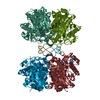
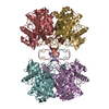
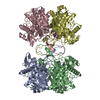
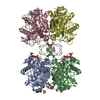
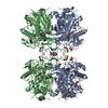
| Title | Crystal structure of MSM-13, a putative T1-like thiolase from Mycobacterium smegmatis | ||||||
 Components Components | Beta-ketothiolase | ||||||
 Keywords Keywords | TRANSFERASE / thiolase / mycobacterium smegmatis / T1-like / tetrameric | ||||||
| Function / homology |  Function and homology information Function and homology informationacetyl-CoA C-acetyltransferase / acetyl-CoA C-acetyltransferase activity Similarity search - Function | ||||||
| Biological species |  Mycobacterium smegmatis str. MC2 155 (bacteria) Mycobacterium smegmatis str. MC2 155 (bacteria) | ||||||
| Method |  X-RAY DIFFRACTION / SYNCHROTRON / MOLECULAR REPLACEMENT / Resolution: 2.162 Å X-RAY DIFFRACTION / SYNCHROTRON / MOLECULAR REPLACEMENT / Resolution: 2.162 Å | ||||||
 Authors Authors | Janardan, N. / Harijan, R.K. / Keima, T.R. / Wierenga, R. / Murthy, M.R.N. | ||||||
 Citation Citation | Journal: Acta Crystallogr.,Sect.D / Year: 2015 Title: Structural characterization of a mitochondrial 3-ketoacyl-CoA (T1)-like thiolase from Mycobacterium smegmatis Authors: Janardan, N. / Harijan, R.K. / Kiema, T.R. / Wierenga, R.K. / Murthy, M.R. | ||||||
| History |
|
- Structure visualization
| Structure viewer | Molecule: MolmilJmol/JSmol |
|---|
- Downloads & links
-Download
| PDBx/mmCIF format | 5byv.cif.gz | 874.7 KB | Display | PDBx/mmCIF format |
|---|---|---|---|---|
| PDB format | pdb5byv.ent.gz | 730.2 KB | Display | PDB format |
| PDBx/mmJSON format | 5byv.json.gz | Tree view | PDBx/mmJSON format | |
| Others |  Other downloads Other downloads |
-Validation report
| Arichive directory | https://data.pdbj.org/pub/pdb/validation_reports/by/5byvftp://data.pdbj.org/pub/pdb/validation_reports/by/5byv | HTTPS FTP |
|---|
-Related structure data
| Related structure data | 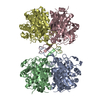 4zrcC 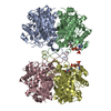 5bz4C 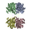 5cbqC 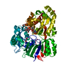 4zecS C: citing same article ( S: Starting model for refinement |
|---|---|
| Similar structure data |


-Links
 PDBj
PDBj

- Assembly
| Deposited unit | 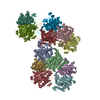
| ||||||||
|---|---|---|---|---|---|---|---|---|---|
| 1 | 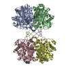
| ||||||||
| 2 | 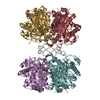
| ||||||||
| 3 | 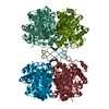
| ||||||||
| Unit cell |
|
-Components
| #1: Protein | Mass: 43007.824 Da / Num. of mol.: 12 Source method: isolated from a genetically manipulated source Source: (gene. exp.) Mycobacterium smegmatis str. MC2 155 (bacteria)Strain: MC2 155 / Gene: MSMEG_2207 / Production host: #2: Water | ChemComp-HOH / |  Mass: 18.015 Da / Num. of mol.: 1526 / Source method: isolated from a natural source / Formula: H2O Mass: 18.015 Da / Num. of mol.: 1526 / Source method: isolated from a natural source / Formula: H2O |
|---|
-Experimental details
-Experiment
| Experiment | Method: X-RAY DIFFRACTION / Number of used crystals: 1 |
|---|
- Sample preparation
| Crystal | Density Matthews: 2.7 Å3/Da / Density % sol: 54.43 % |
|---|---|
| Crystal grow | Temperature: 293 K / Method: microbatch / pH: 6 Details: 45% PEG 200, 0.1M MES monohydrate pH 6.0, 0.07M calcium chloride dihydrate |
-Data collection
| Diffraction | Mean temperature: 100 K |
|---|---|
| Diffraction source | Source: SYNCHROTRON / Site: ESRF  / Beamline: BM14 / Wavelength: 0.95 Å / Beamline: BM14 / Wavelength: 0.95 Å |
| Detector | Type: MAR scanner 300 mm plate / Detector: IMAGE PLATE / Date: Jul 8, 2014 |
| Radiation | Protocol: SINGLE WAVELENGTH / Monochromatic (M) / Laue (L): M / Scattering type: x-ray |
| Radiation wavelength | Wavelength: 0.95 Å / Relative weight: 1 |
| Reflection | Resolution: 2.16→45.193 Å / Num. obs: 290242 / % possible obs: 99.94 % / Redundancy: 1.9 % / Net I/σ(I): 12.2 |
- Processing
| Software |
| |||||||||||||||||||||||||||||||||||||||||||||||||||||||||||||||||||||||||||||||||||||||||||||||||||||||||||||||||||||||||||||||||||||||||||||||||||
|---|---|---|---|---|---|---|---|---|---|---|---|---|---|---|---|---|---|---|---|---|---|---|---|---|---|---|---|---|---|---|---|---|---|---|---|---|---|---|---|---|---|---|---|---|---|---|---|---|---|---|---|---|---|---|---|---|---|---|---|---|---|---|---|---|---|---|---|---|---|---|---|---|---|---|---|---|---|---|---|---|---|---|---|---|---|---|---|---|---|---|---|---|---|---|---|---|---|---|---|---|---|---|---|---|---|---|---|---|---|---|---|---|---|---|---|---|---|---|---|---|---|---|---|---|---|---|---|---|---|---|---|---|---|---|---|---|---|---|---|---|---|---|---|---|---|---|---|---|
| Refinement | Method to determine structure: MOLECULAR REPLACEMENT Starting model: 4ZEC Resolution: 2.162→45.193 Å / Cross valid method: FREE R-VALUE / σ(F): 0.2 / Phase error: 24.35 / Stereochemistry target values: TWIN_LSQ_F
| |||||||||||||||||||||||||||||||||||||||||||||||||||||||||||||||||||||||||||||||||||||||||||||||||||||||||||||||||||||||||||||||||||||||||||||||||||
| Solvent computation | Shrinkage radii: 0.9 Å / VDW probe radii: 1.11 Å / Solvent model: FLAT BULK SOLVENT MODEL | |||||||||||||||||||||||||||||||||||||||||||||||||||||||||||||||||||||||||||||||||||||||||||||||||||||||||||||||||||||||||||||||||||||||||||||||||||
| Refinement step | Cycle: LAST / Resolution: 2.162→45.193 Å
| |||||||||||||||||||||||||||||||||||||||||||||||||||||||||||||||||||||||||||||||||||||||||||||||||||||||||||||||||||||||||||||||||||||||||||||||||||
| Refine LS restraints |
| |||||||||||||||||||||||||||||||||||||||||||||||||||||||||||||||||||||||||||||||||||||||||||||||||||||||||||||||||||||||||||||||||||||||||||||||||||
| LS refinement shell |
|
