 Movie
Movie Controller

[English] 日本語
 Yorodumi
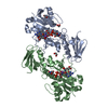
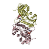
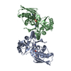
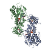
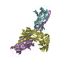
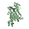
- PDB-7n7b: crystal structure of the N-formyltrasferase HCAN_0200 from Helico... -
+ Open data

- Basic information
| Entry | Database: PDB / ID: 7n7b | ||||||
|---|---|---|---|---|---|---|---|
| Title | crystal structure of the N-formyltrasferase HCAN_0200 from Helicobacter canadensis on complex with folinic acid and dTDP-3-aminofucose | ||||||
 Components Components | Formyl_trans_N domain-containing protein | ||||||
 Keywords Keywords | TRANSFERASE / folinic acid / formyltransferase / aminofucose | ||||||
| Function / homology | methionyl-tRNA formyltransferase activity / Formyl transferase, N-terminal / Formyl transferase / Formyl transferase, N-terminal domain superfamily / Chem-FON / Chem-T3F / Formyl transferase N-terminal domain-containing protein Function and homology information Function and homology information | ||||||
| Biological species |  Helicobacter canadensis MIT 98-5491 (bacteria) Helicobacter canadensis MIT 98-5491 (bacteria) | ||||||
| Method |  X-RAY DIFFRACTION / FOURIER SYNTHESIS / Resolution: 2 Å X-RAY DIFFRACTION / FOURIER SYNTHESIS / Resolution: 2 Å | ||||||
 Authors Authors | Heisdorf, C.J. / Thoden, J.B. / Holden, H.M. | ||||||
| Funding support |  United States, 1items United States, 1items
| ||||||
 Citation Citation | Journal: Protein Sci. / Year: 2021 Title: Investigation of the enzymes required for the biosynthesis of an unusual formylated sugar in the emerging human pathogen Helicobacter canadensis. Authors: Heisdorf, C.J. / Griffiths, W.A. / Thoden, J.B. / Holden, H.M. | ||||||
| History |
|
- Structure visualization
| Structure viewer | Molecule: MolmilJmol/JSmol |
|---|
- Downloads & links
-Download
| PDBx/mmCIF format | 7n7b.cif.gz | 141.3 KB | Display | PDBx/mmCIF format |
|---|---|---|---|---|
| PDB format | pdb7n7b.ent.gz | 107.4 KB | Display | PDB format |
| PDBx/mmJSON format | 7n7b.json.gz | Tree view | PDBx/mmJSON format | |
| Others |  Other downloads Other downloads |
-Validation report
| Arichive directory | https://data.pdbj.org/pub/pdb/validation_reports/n7/7n7bftp://data.pdbj.org/pub/pdb/validation_reports/n7/7n7b | HTTPS FTP |
|---|
-Related structure data
| Related structure data |  7n63C 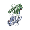 7n67C 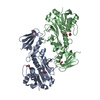 7n7aSC  7n7cC S: Starting model for refinement C: citing same article ( |
|---|---|
| Similar structure data |


-Links
 PDBj
PDBj- Assembly
| Deposited unit | 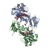
| ||||||||
|---|---|---|---|---|---|---|---|---|---|
| 1 |
| ||||||||
| Unit cell |
|
-Components
-Protein , 1 types, 2 molecules AB
| #1: Protein | Mass: 33065.656 Da / Num. of mol.: 2 Source method: isolated from a genetically manipulated source Source: (gene. exp.) Helicobacter canadensis MIT 98-5491 (bacteria)Gene: HCAN_0200 / Production host: |
|---|
-Non-polymers , 5 types, 464 molecules 
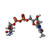


| #2: Chemical |  Mass: 473.439 Da / Num. of mol.: 2 / Source method: obtained synthetically / Formula: C20H23N7O7 / Feature type: SUBJECT OF INVESTIGATION Mass: 473.439 Da / Num. of mol.: 2 / Source method: obtained synthetically / Formula: C20H23N7O7 / Feature type: SUBJECT OF INVESTIGATION#3: Chemical |  Mass: 547.345 Da / Num. of mol.: 2 / Source method: obtained synthetically / Formula: C16H27N3O14P2 / Feature type: SUBJECT OF INVESTIGATION Mass: 547.345 Da / Num. of mol.: 2 / Source method: obtained synthetically / Formula: C16H27N3O14P2 / Feature type: SUBJECT OF INVESTIGATION#4: Chemical | ChemComp-MES / |  Mass: 195.237 Da / Num. of mol.: 1 / Source method: obtained synthetically / Formula: C6H13NO4S / Comment: pH buffer*YM Mass: 195.237 Da / Num. of mol.: 1 / Source method: obtained synthetically / Formula: C6H13NO4S / Comment: pH buffer*YM#5: Chemical | ChemComp-EDO /  Mass: 62.068 Da / Num. of mol.: 5 / Source method: obtained synthetically / Formula: C2H6O2 Mass: 62.068 Da / Num. of mol.: 5 / Source method: obtained synthetically / Formula: C2H6O2#6: Water | ChemComp-HOH / | Mass: 18.015 Da / Num. of mol.: 454 / Source method: isolated from a natural source / Formula: H2O |
|---|
-Details
| Has ligand of interest | Y |
|---|
-Experimental details
-Experiment
| Experiment | Method: X-RAY DIFFRACTION / Number of used crystals: 1 |
|---|
- Sample preparation
| Crystal | Density Matthews: 2.68 Å3/Da / Density % sol: 54.08 % |
|---|---|
| Crystal grow | Temperature: 293 K / Method: vapor diffusion, hanging drop / pH: 5 Details: 11-14% PEG-8000, 2% DMSO, 100 mM homopipes (pH 5). Crystals soaked in 26% PEG-8000, 600 mM NaCl, 5 mM folinic acis, 10 mM dTDP-Fuc3N, 100 mM MES (pH 6) |
-Data collection
| Diffraction | Mean temperature: 100 K / Serial crystal experiment: N |
|---|---|
| Diffraction source | Source: SEALED TUBE / Type: BRUKER D8 QUEST / Wavelength: 1.5418 Å |
| Detector | Type: Bruker PHOTON II / Detector: PIXEL / Date: Oct 18, 2019 |
| Radiation | Protocol: SINGLE WAVELENGTH / Monochromatic (M) / Laue (L): M / Scattering type: x-ray |
| Radiation wavelength | Wavelength: 1.5418 Å / Relative weight: 1 |
| Reflection | Resolution: 2→50 Å / Num. obs: 45715 / % possible obs: 96.6 % / Observed criterion σ(F): 0 / Observed criterion σ(I): 0 / Redundancy: 5.6 % / Rsym value: 0.074 / Net I/σ(I): 13.5 |
| Reflection shell | Resolution: 2→2.1 Å / Redundancy: 3.5 % / Mean I/σ(I) obs: 2.8 / Num. unique obs: 5967 / Rsym value: 0.334 / % possible all: 93 |
- Processing
| Software |
| ||||||||||||||||||||||||||||||||||||||||||||||||||||||||||||
|---|---|---|---|---|---|---|---|---|---|---|---|---|---|---|---|---|---|---|---|---|---|---|---|---|---|---|---|---|---|---|---|---|---|---|---|---|---|---|---|---|---|---|---|---|---|---|---|---|---|---|---|---|---|---|---|---|---|---|---|---|---|
| Refinement | Method to determine structure: FOURIER SYNTHESIS Starting model: 7n7a Resolution: 2→29.64 Å / Cor.coef. Fo:Fc: 0.952 / Cor.coef. Fo:Fc free: 0.906 / SU B: 5.184 / SU ML: 0.141 / Cross valid method: THROUGHOUT / σ(F): 0 / ESU R: 0.187 / ESU R Free: 0.179 / Stereochemistry target values: MAXIMUM LIKELIHOOD Details: HYDROGENS HAVE BEEN ADDED IN THE RIDING POSITIONS U VALUES : REFINED INDIVIDUALLY
| ||||||||||||||||||||||||||||||||||||||||||||||||||||||||||||
| Solvent computation | Ion probe radii: 0.8 Å / Shrinkage radii: 0.8 Å / VDW probe radii: 1.2 Å / Solvent model: MASK | ||||||||||||||||||||||||||||||||||||||||||||||||||||||||||||
| Displacement parameters | Biso max: 86.33 Å2 / Biso mean: 30.505 Å2 / Biso min: 11.44 Å2
| ||||||||||||||||||||||||||||||||||||||||||||||||||||||||||||
| Refinement step | Cycle: final / Resolution: 2→29.64 Å
| ||||||||||||||||||||||||||||||||||||||||||||||||||||||||||||
| Refine LS restraints |
| ||||||||||||||||||||||||||||||||||||||||||||||||||||||||||||
| LS refinement shell | Resolution: 2→2.052 Å / Rfactor Rfree error: 0 / Total num. of bins used: 20
|
