 Movie
Movie Controller

[English] 日本語
 Yorodumi
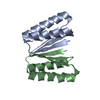
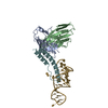
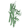
- PDB-7mwq: Structure of De Novo designed beta sheet heterodimer LHD29A53/B53 -
+ Open data

- Basic information
| Entry | Database: PDB / ID: 7mwq | ||||||
|---|---|---|---|---|---|---|---|
| Title | Structure of De Novo designed beta sheet heterodimer LHD29A53/B53 | ||||||
 Components Components |
| ||||||
 Keywords Keywords | DE NOVO PROTEIN / de novo design / beta-sheet / heterodimer | ||||||
| Biological species | synthetic construct (others) | ||||||
| Method |  X-RAY DIFFRACTION / SYNCHROTRON / MOLECULAR REPLACEMENT / Resolution: 2.56 Å X-RAY DIFFRACTION / SYNCHROTRON / MOLECULAR REPLACEMENT / Resolution: 2.56 Å | ||||||
 Authors Authors | Bera, A.K. / Sahtoe, D.D. / Kang, A. / Praetorius, F. / Baker, D. | ||||||
| Funding support |  United States, 1items United States, 1items
| ||||||
 Citation Citation | Journal: Science / Year: 2022 Title: Reconfigurable asymmetric protein assemblies through implicit negative design. Authors: Sahtoe, D.D. / Praetorius, F. / Courbet, A. / Hsia, Y. / Wicky, B.I.M. / Edman, N.I. / Miller, L.M. / Timmermans, B.J.R. / Decarreau, J. / Morris, H.M. / Kang, A. / Bera, A.K. / Baker, D. | ||||||
| History |
|
- Structure visualization
| Structure viewer | Molecule:  MolmilJmol/JSmol MolmilJmol/JSmol |
|---|
- Downloads & links
-Download
| PDBx/mmCIF format | 7mwq.cif.gz | 202 KB | Display | PDBx/mmCIF format |
|---|---|---|---|---|
| PDB format | pdb7mwq.ent.gz | 130.8 KB | Display | PDB format |
| PDBx/mmJSON format | 7mwq.json.gz | Tree view | PDBx/mmJSON format | |
| Others |  Other downloads Other downloads |
-Validation report
| Arichive directory | https://data.pdbj.org/pub/pdb/validation_reports/mw/7mwqftp://data.pdbj.org/pub/pdb/validation_reports/mw/7mwq | HTTPS FTP |
|---|
-Related structure data


-Links
 PDBj
PDBj
- Assembly
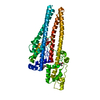
| Deposited unit | 
| ||||||||||||
|---|---|---|---|---|---|---|---|---|---|---|---|---|---|
| 1 | 
| ||||||||||||
| 2 | 
| ||||||||||||
| Unit cell |
|
-Components
| #1: Protein | Mass: 22983.434 Da / Num. of mol.: 2 Source method: isolated from a genetically manipulated source Source: (gene. exp.) synthetic construct (others) / Production host:  #2: Protein | Mass: 23192.820 Da / Num. of mol.: 2 Source method: isolated from a genetically manipulated source Source: (gene. exp.) synthetic construct (others) / Production host: #3: Water | ChemComp-HOH / |  Mass: 18.015 Da / Num. of mol.: 14 / Source method: isolated from a natural source / Formula: H2O Mass: 18.015 Da / Num. of mol.: 14 / Source method: isolated from a natural source / Formula: H2O |
|---|
-Experimental details
-Experiment
| Experiment | Method: X-RAY DIFFRACTION / Number of used crystals: 1 |
|---|
- Sample preparation
| Crystal | Density Matthews: 2.94 Å3/Da / Density % sol: 58.11 % |
|---|---|
| Crystal grow | Temperature: 293 K / Method: vapor diffusion, sitting drop / Details: 0.2M Sodium Iodide, 20% PEG3350 |
-Data collection
| Diffraction | Mean temperature: 100 K / Serial crystal experiment: N |
|---|---|
| Diffraction source | Source: SYNCHROTRON / Site: ALS / Beamline: 8.2.1 / Wavelength: 0.99991 Å |
| Detector | Type: ADSC QUANTUM 315r / Detector: CCD / Date: Feb 25, 2020 |
| Radiation | Protocol: SINGLE WAVELENGTH / Monochromatic (M) / Laue (L): M / Scattering type: x-ray |
| Radiation wavelength | Wavelength: 0.99991 Å / Relative weight: 1 |
| Reflection | Resolution: 2.56→51.56 Å / Num. obs: 32600 / % possible obs: 97.9 % / Redundancy: 2.3 % / Biso Wilson estimate: 58.44 Å2 / CC1/2: 0.991 / Net I/σ(I): 4.7 |
| Reflection shell | Resolution: 2.56→2.65 Å / Redundancy: 2.4 % / Mean I/σ(I) obs: 1.1 / Num. unique obs: 2389 / CC1/2: 0.651 / % possible all: 93.4 |
- Processing
| Software |
| |||||||||||||||||||||||||||||||||||||||||||||||||||||||||||||||||||||||||||||||||||||||||||
|---|---|---|---|---|---|---|---|---|---|---|---|---|---|---|---|---|---|---|---|---|---|---|---|---|---|---|---|---|---|---|---|---|---|---|---|---|---|---|---|---|---|---|---|---|---|---|---|---|---|---|---|---|---|---|---|---|---|---|---|---|---|---|---|---|---|---|---|---|---|---|---|---|---|---|---|---|---|---|---|---|---|---|---|---|---|---|---|---|---|---|---|---|
| Refinement | Method to determine structure: MOLECULAR REPLACEMENT Starting model: Design model Resolution: 2.56→51.56 Å / SU ML: 0.4518 / Cross valid method: FREE R-VALUE / σ(F): 1.96 / Phase error: 32.5726 Stereochemistry target values: GeoStd + Monomer Library + CDL v1.2
| |||||||||||||||||||||||||||||||||||||||||||||||||||||||||||||||||||||||||||||||||||||||||||
| Solvent computation | Shrinkage radii: 0.9 Å / VDW probe radii: 1.11 Å / Solvent model: FLAT BULK SOLVENT MODEL | |||||||||||||||||||||||||||||||||||||||||||||||||||||||||||||||||||||||||||||||||||||||||||
| Displacement parameters | Biso mean: 75.6 Å2 | |||||||||||||||||||||||||||||||||||||||||||||||||||||||||||||||||||||||||||||||||||||||||||
| Refinement step | Cycle: LAST / Resolution: 2.56→51.56 Å
| |||||||||||||||||||||||||||||||||||||||||||||||||||||||||||||||||||||||||||||||||||||||||||
| Refine LS restraints |
| |||||||||||||||||||||||||||||||||||||||||||||||||||||||||||||||||||||||||||||||||||||||||||
| LS refinement shell |
|
