 Movie
Movie Controller

[English] 日本語
 Yorodumi
- PDB-7mq2: C9A Streptococcus pneumoniae CstR in the reduced state, space gro... -
+ Open data

- Basic information
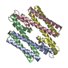
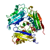
| Entry | Database: PDB / ID: 7mq2 | ||||||
|---|---|---|---|---|---|---|---|
| Title | C9A Streptococcus pneumoniae CstR in the reduced state, space group P21 | ||||||
 Components Components | Copper-sensing transcriptional repressor csoR | ||||||
 Keywords Keywords | TRANSCRIPTION / transcriptional regulator / persulfide sensor / CsoR family / CstR family | ||||||
| Function / homology | Metal-sensitive transcriptional repressor / Metal-sensitive repressor, helix protomer superfamily / Metal-sensitive transcriptional repressor / negative regulation of DNA-templated transcription / DNA binding / metal ion binding / Copper-sensing transcriptional repressor csoR Function and homology information Function and homology information | ||||||
| Biological species |  Streptococcus pneumoniae D39 (bacteria) Streptococcus pneumoniae D39 (bacteria) | ||||||
| Method |  X-RAY DIFFRACTION / SYNCHROTRON / SAD / Resolution: 2.29 Å X-RAY DIFFRACTION / SYNCHROTRON / SAD / Resolution: 2.29 Å | ||||||
 Authors Authors | Fakhoury, J.N. / Gonzalez-Gutierrez, G. / Giedroc, D.P. | ||||||
| Funding support |  United States, 1items United States, 1items
| ||||||
 Citation Citation | Journal: Nucleic Acids Res. / Year: 2021 Title: Functional asymmetry and chemical reactivity of CsoR family persulfide sensors. Authors: Fakhoury, J.N. / Zhang, Y. / Edmonds, K.A. / Bringas, M. / Luebke, J.L. / Gonzalez-Gutierrez, G. / Capdevila, D.A. / Giedroc, D.P. | ||||||
| History |
|
- Structure visualization
| Structure viewer | Molecule: MolmilJmol/JSmol |
|---|
- Downloads & links
-Download
| PDBx/mmCIF format | 7mq2.cif.gz | 153.7 KB | Display | PDBx/mmCIF format |
|---|---|---|---|---|
| PDB format | pdb7mq2.ent.gz | 103.1 KB | Display | PDB format |
| PDBx/mmJSON format | 7mq2.json.gz | Tree view | PDBx/mmJSON format | |
| Others |  Other downloads Other downloads |
-Validation report
| Summary document | 7mq2_validation.pdf.gz | 455.2 KB | Display | wwPDB validaton report |
|---|---|---|---|---|
| Full document | 7mq2_full_validation.pdf.gz | 458.9 KB | Display | |
| Data in XML | 7mq2_validation.xml.gz | 12.3 KB | Display | |
| Data in CIF | 7mq2_validation.cif.gz | 16.6 KB | Display | |
| Arichive directory | https://data.pdbj.org/pub/pdb/validation_reports/mq/7mq2ftp://data.pdbj.org/pub/pdb/validation_reports/mq/7mq2 | HTTPS FTP |
-Related structure data


-Links
 PDBj
PDBj- Assembly
| Deposited unit | 
| ||||||||||||
|---|---|---|---|---|---|---|---|---|---|---|---|---|---|
| 1 |
| ||||||||||||
| Unit cell |
|
-Components
| #1: Protein | Mass: 9831.981 Da / Num. of mol.: 4 / Mutation: C9A Source method: isolated from a genetically manipulated source Source: (gene. exp.) Streptococcus pneumoniae D39 (bacteria)Gene: csoR, ERS019420_01408, GM542_04805, SAMEA2335968_01957 Production host: #2: Water | ChemComp-HOH / |  Mass: 18.015 Da / Num. of mol.: 21 / Source method: isolated from a natural source / Formula: H2O Mass: 18.015 Da / Num. of mol.: 21 / Source method: isolated from a natural source / Formula: H2OHas ligand of interest | N | Has protein modification | Y | |
|---|
-Experimental details
-Experiment
| Experiment | Method: X-RAY DIFFRACTION / Number of used crystals: 1 |
|---|
- Sample preparation
| Crystal | Density Matthews: 2.08 Å3/Da / Density % sol: 40.9 % |
|---|---|
| Crystal grow | Temperature: 293 K / Method: vapor diffusion, sitting drop / pH: 7.5 / Details: 0.1 M HEPES pH 7.5, PEG 200 34-40% |
-Data collection
| Diffraction | Mean temperature: 100 K / Serial crystal experiment: N |
|---|---|
| Diffraction source | Source: SYNCHROTRON / Site: ALS / Beamline: 4.2.2 / Wavelength: 0.97625 Å |
| Detector | Type: RDI CMOS_8M / Detector: CMOS / Date: Sep 26, 2019 |
| Radiation | Protocol: SINGLE WAVELENGTH / Monochromatic (M) / Laue (L): M / Scattering type: x-ray |
| Radiation wavelength | Wavelength: 0.97625 Å / Relative weight: 1 |
| Reflection | Resolution: 2.29→45.57 Å / Num. obs: 14665 / % possible obs: 99.9 % / Redundancy: 7 % / Biso Wilson estimate: 48.39 Å2 / CC1/2: 0.999 / Rpim(I) all: 0.028 / Rrim(I) all: 0.074 / Rsym value: 0.069 / Net I/σ(I): 16.2 |
| Reflection shell | Resolution: 2.29→2.37 Å / Redundancy: 7 % / Mean I/σ(I) obs: 1.7 / Num. unique obs: 1408 / CC1/2: 0.811 / Rpim(I) all: 0.374 / Rrim(I) all: 0.988 / Rsym value: 0.925 / % possible all: 100 |
- Processing
| Software |
| ||||||||||||||||||||||||||||||||||||||||||||||||||||||||||||||||||||||||||||||||||||
|---|---|---|---|---|---|---|---|---|---|---|---|---|---|---|---|---|---|---|---|---|---|---|---|---|---|---|---|---|---|---|---|---|---|---|---|---|---|---|---|---|---|---|---|---|---|---|---|---|---|---|---|---|---|---|---|---|---|---|---|---|---|---|---|---|---|---|---|---|---|---|---|---|---|---|---|---|---|---|---|---|---|---|---|---|---|
| Refinement | Method to determine structure: SAD / Resolution: 2.29→36.26 Å / SU ML: 0.3516 / Cross valid method: FREE R-VALUE / σ(F): 1.24 / Phase error: 34.8125 Stereochemistry target values: GeoStd + Monomer Library + CDL v1.2
| ||||||||||||||||||||||||||||||||||||||||||||||||||||||||||||||||||||||||||||||||||||
| Solvent computation | Shrinkage radii: 0.9 Å / VDW probe radii: 1.11 Å / Solvent model: FLAT BULK SOLVENT MODEL | ||||||||||||||||||||||||||||||||||||||||||||||||||||||||||||||||||||||||||||||||||||
| Displacement parameters | Biso mean: 75.24 Å2 | ||||||||||||||||||||||||||||||||||||||||||||||||||||||||||||||||||||||||||||||||||||
| Refinement step | Cycle: LAST / Resolution: 2.29→36.26 Å
| ||||||||||||||||||||||||||||||||||||||||||||||||||||||||||||||||||||||||||||||||||||
| Refine LS restraints |
| ||||||||||||||||||||||||||||||||||||||||||||||||||||||||||||||||||||||||||||||||||||
| LS refinement shell |
| ||||||||||||||||||||||||||||||||||||||||||||||||||||||||||||||||||||||||||||||||||||
| Refinement TLS params. | Method: refined / Origin x: -7.94626244153 Å / Origin y: 18.572731771 Å / Origin z: -14.1342028241 Å
| ||||||||||||||||||||||||||||||||||||||||||||||||||||||||||||||||||||||||||||||||||||
| Refinement TLS group | Selection details: all |
