 Movie
Movie Controller


+ Open data

- Basic information
| Entry | Database: PDB / ID: 7mon | ||||||
|---|---|---|---|---|---|---|---|
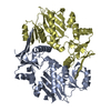
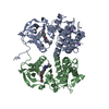
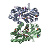
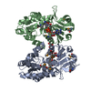
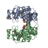
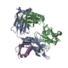
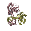
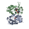
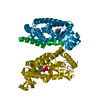
| Title | Structure of human RIPK3-MLKL complex | ||||||
 Components Components |
| ||||||
 Keywords Keywords | TRANSFERASE / pseudokinase / kinase / complex / necroptosis | ||||||
| Function / homology |  Function and homology information Function and homology informationregulation of activation-induced cell death of T cells / regulation of CD8-positive, alpha-beta cytotoxic T cell extravasation / execution phase of necroptosis / regulation of T cell mediated cytotoxicity / ripoptosome assembly involved in necroptotic process / regulation of adaptive immune response / regulation of activated T cell proliferation / programmed necrotic cell death / Microbial modulation of RIPK1-mediated regulated necrosis / ripoptosome ...regulation of activation-induced cell death of T cells / regulation of CD8-positive, alpha-beta cytotoxic T cell extravasation / execution phase of necroptosis / regulation of T cell mediated cytotoxicity / ripoptosome assembly involved in necroptotic process / regulation of adaptive immune response / regulation of activated T cell proliferation / programmed necrotic cell death / Microbial modulation of RIPK1-mediated regulated necrosis / ripoptosome / TRIF-mediated programmed cell death / regulation of type II interferon production / TLR3-mediated TICAM1-dependent programmed cell death / activation of protein kinase activity / SARS-CoV-1-mediated effects on programmed cell death / necroptotic signaling pathway / RIP-mediated NFkB activation via ZBP1 / positive regulation of necroptotic process / RIPK1-mediated regulated necrosis / non-canonical NF-kappaB signal transduction / necroptotic process / protein homotrimerization / T cell homeostasis / TRP channels / lymph node development / spleen development / positive regulation of intrinsic apoptotic signaling pathway / : / reactive oxygen species metabolic process / thymus development / protein modification process / TICAM1, RIP1-mediated IKK complex recruitment / protein serine/threonine kinase binding / IKK complex recruitment mediated by RIP1 / apoptotic signaling pathway / Regulation of necroptotic cell death / positive regulation of reactive oxygen species metabolic process / cellular response to hydrogen peroxide / cell junction / SARS-CoV-1 activates/modulates innate immune responses / T cell differentiation in thymus / regulation of apoptotic process / defense response to virus / amyloid fibril formation / protein kinase activity / transcription coactivator activity / non-specific serine/threonine protein kinase / cell surface receptor signaling pathway / protein serine kinase activity / protein serine/threonine kinase activity / protein kinase binding / protein-containing complex binding / signal transduction / protein-containing complex / ATP binding / identical protein binding / nucleus / plasma membrane / cytosol / cytoplasm Similarity search - Function | ||||||
| Biological species |  Homo sapiens (human) Homo sapiens (human) | ||||||
| Method |  X-RAY DIFFRACTION / SYNCHROTRON / MOLECULAR REPLACEMENT / molecular replacement / Resolution: 2.23 Å X-RAY DIFFRACTION / SYNCHROTRON / MOLECULAR REPLACEMENT / molecular replacement / Resolution: 2.23 Å | ||||||
 Authors Authors | Meng, Y. / Davies, K.A. / Czabotar, P.E. / Murphy, J.M. | ||||||
| Funding support |  Australia, 1items Australia, 1items
| ||||||
 Citation Citation | Journal: Nat Commun / Year: 2021 Title: Human RIPK3 maintains MLKL in an inactive conformation prior to cell death by necroptosis. Authors: Meng, Y. / Davies, K.A. / Fitzgibbon, C. / Young, S.N. / Garnish, S.E. / Horne, C.R. / Luo, C. / Garnier, J.M. / Liang, L.Y. / Cowan, A.D. / Samson, A.L. / Lessene, G. / Sandow, J.J. / ...Authors: Meng, Y. / Davies, K.A. / Fitzgibbon, C. / Young, S.N. / Garnish, S.E. / Horne, C.R. / Luo, C. / Garnier, J.M. / Liang, L.Y. / Cowan, A.D. / Samson, A.L. / Lessene, G. / Sandow, J.J. / Czabotar, P.E. / Murphy, J.M. | ||||||
| History |
|
- Structure visualization
| Structure viewer | Molecule: MolmilJmol/JSmol |
|---|
- Downloads & links
-Download
| PDBx/mmCIF format | 7mon.cif.gz | 130.1 KB | Display | PDBx/mmCIF format |
|---|---|---|---|---|
| PDB format | pdb7mon.ent.gz | 97 KB | Display | PDB format |
| PDBx/mmJSON format | 7mon.json.gz | Tree view | PDBx/mmJSON format | |
| Others |  Other downloads Other downloads |
-Validation report
| Arichive directory | https://data.pdbj.org/pub/pdb/validation_reports/mo/7monftp://data.pdbj.org/pub/pdb/validation_reports/mo/7mon | HTTPS FTP |
|---|
-Related structure data
| Related structure data | 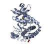 7mx3C 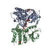 4m69S 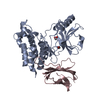 7jxuS S: Starting model for refinement C: citing same article ( |
|---|---|
| Similar structure data |

-Links
 PDBj
PDBj


- Assembly
| Deposited unit | 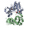
| ||||||||
|---|---|---|---|---|---|---|---|---|---|
| 1 |
| ||||||||
| Unit cell |
|
-Components
| #1: Protein | Mass: 32736.750 Da / Num. of mol.: 1 Source method: isolated from a genetically manipulated source Source: (gene. exp.) Homo sapiens (human) / Gene: MLKL / Production host:   Spodoptera frugiperda (fall armyworm) / References: UniProt: Q8NB16 Spodoptera frugiperda (fall armyworm) / References: UniProt: Q8NB16 | ||||||
|---|---|---|---|---|---|---|---|
| #2: Protein | Mass: 35064.777 Da / Num. of mol.: 1 / Mutation: C3S, C110A Source method: isolated from a genetically manipulated source Source: (gene. exp.) Homo sapiens (human) / Gene: RIPK3, RIP3 / Production host: Spodoptera frugiperda (fall armyworm)References: UniProt: Q9Y572, non-specific serine/threonine protein kinase | ||||||
| #3: Chemical |   Mass: 502.469 Da / Num. of mol.: 2 / Source method: obtained synthetically / Formula: C27H20F2N4O4 / Feature type: SUBJECT OF INVESTIGATION Mass: 502.469 Da / Num. of mol.: 2 / Source method: obtained synthetically / Formula: C27H20F2N4O4 / Feature type: SUBJECT OF INVESTIGATION#4: Water | ChemComp-HOH / |  Mass: 18.015 Da / Num. of mol.: 105 / Source method: isolated from a natural source / Formula: H2O Mass: 18.015 Da / Num. of mol.: 105 / Source method: isolated from a natural source / Formula: H2OHas ligand of interest | Y | Has protein modification | Y | |
-Experimental details
-Experiment
| Experiment | Method: X-RAY DIFFRACTION / Number of used crystals: 1 |
|---|
- Sample preparation
| Crystal | Density Matthews: 2.07 Å3/Da / Density % sol: 40.45 % |
|---|---|
| Crystal grow | Temperature: 281.15 K / Method: vapor diffusion, hanging drop / pH: 5.5 Details: 0.2 M ammonium acetate 30%w/v PEG 4000 0.1 M trisodium citrate-citric acid pH 5.5 |
-Data collection
| Diffraction | Mean temperature: 100 K / Serial crystal experiment: N | ||||||||||||||||||||||||||||||
|---|---|---|---|---|---|---|---|---|---|---|---|---|---|---|---|---|---|---|---|---|---|---|---|---|---|---|---|---|---|---|---|
| Diffraction source | Source: SYNCHROTRON / Site: Australian Synchrotron / Beamline: MX2 / Wavelength: 0.9537 Å | ||||||||||||||||||||||||||||||
| Detector | Type: DECTRIS EIGER X 16M / Detector: PIXEL / Date: Feb 11, 2021 | ||||||||||||||||||||||||||||||
| Radiation | Protocol: SINGLE WAVELENGTH / Monochromatic (M) / Laue (L): M / Scattering type: x-ray | ||||||||||||||||||||||||||||||
| Radiation wavelength | Wavelength: 0.9537 Å / Relative weight: 1 | ||||||||||||||||||||||||||||||
| Reflection | Resolution: 2.23→45.78 Å / Num. obs: 28056 / % possible obs: 99.4 % / Redundancy: 6.7 % / CC1/2: 0.997 / Rmerge(I) obs: 0.144 / Rpim(I) all: 0.06 / Rrim(I) all: 0.156 / Net I/σ(I): 8.3 / Num. measured all: 186701 / Scaling rejects: 17 | ||||||||||||||||||||||||||||||
| Reflection shell | Diffraction-ID: 1
|
-Phasing
| Phasing | Method: molecular replacement | |||||||||
|---|---|---|---|---|---|---|---|---|---|---|
| Phasing MR |
|
- Processing
| Software |
| |||||||||||||||||||||||||||||||||||||||||||||||||||||||||||||||||||||||||||||||||||||||||||||||||||||||||
|---|---|---|---|---|---|---|---|---|---|---|---|---|---|---|---|---|---|---|---|---|---|---|---|---|---|---|---|---|---|---|---|---|---|---|---|---|---|---|---|---|---|---|---|---|---|---|---|---|---|---|---|---|---|---|---|---|---|---|---|---|---|---|---|---|---|---|---|---|---|---|---|---|---|---|---|---|---|---|---|---|---|---|---|---|---|---|---|---|---|---|---|---|---|---|---|---|---|---|---|---|---|---|---|---|---|---|
| Refinement | Method to determine structure: MOLECULAR REPLACEMENT Starting model: 7JXU, 4M69 Resolution: 2.23→41.42 Å / SU ML: 0.31 / Cross valid method: THROUGHOUT / σ(F): 1.35 / Phase error: 26.54 / Stereochemistry target values: ML
| |||||||||||||||||||||||||||||||||||||||||||||||||||||||||||||||||||||||||||||||||||||||||||||||||||||||||
| Solvent computation | Shrinkage radii: 0.9 Å / VDW probe radii: 1.11 Å / Solvent model: FLAT BULK SOLVENT MODEL | |||||||||||||||||||||||||||||||||||||||||||||||||||||||||||||||||||||||||||||||||||||||||||||||||||||||||
| Displacement parameters | Biso max: 127.84 Å2 / Biso mean: 43.6036 Å2 / Biso min: 19.96 Å2 | |||||||||||||||||||||||||||||||||||||||||||||||||||||||||||||||||||||||||||||||||||||||||||||||||||||||||
| Refinement step | Cycle: final / Resolution: 2.23→41.42 Å
| |||||||||||||||||||||||||||||||||||||||||||||||||||||||||||||||||||||||||||||||||||||||||||||||||||||||||
| Refine LS restraints |
| |||||||||||||||||||||||||||||||||||||||||||||||||||||||||||||||||||||||||||||||||||||||||||||||||||||||||
| LS refinement shell | Refine-ID: X-RAY DIFFRACTION / Rfactor Rfree error: 0 / Total num. of bins used: 14
|
