 Movie
Movie Controller

[English] 日本語
 Yorodumi
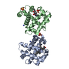
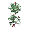
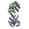
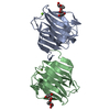
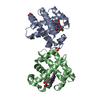
- PDB-7mnh: V59W mutant of Dehaloperoxidase A from Amphitrite ornata treated ... -
+ Open data

- Basic information
| Entry | Database: PDB / ID: 7mnh | ||||||
|---|---|---|---|---|---|---|---|
| Title | V59W mutant of Dehaloperoxidase A from Amphitrite ornata treated with Fluoride | ||||||
 Components Components | Dehaloperoxidase A | ||||||
 Keywords Keywords | OXIDOREDUCTASE / Dehaloperoxidase A / Amphitrite ornata / DHP | ||||||
| Function / homology |  Function and homology information Function and homology informationoxygen carrier activity / peroxidase activity / oxygen binding / heme binding / metal ion binding Similarity search - Function | ||||||
| Biological species |   Amphitrite ornata (invertebrata) Amphitrite ornata (invertebrata) | ||||||
| Method |  X-RAY DIFFRACTION / SYNCHROTRON / MOLECULAR REPLACEMENT / Resolution: 1.24 Å X-RAY DIFFRACTION / SYNCHROTRON / MOLECULAR REPLACEMENT / Resolution: 1.24 Å | ||||||
 Authors Authors | Shay, M.R. / Thompson, M.K. / Franzen, S. | ||||||
 Citation Citation | Journal: J Porphyr Phthalocyanines / Year: 2021 Title: A new inhibition mechanism in the multifunctional catalytic hemoglobin dehaloperoxidase as revealed by the DHP A(V59W) mutant: A spectroscopic and crystallographic study Authors: Thompson, M.K. / Shay, M.R. / Dumarieh, R. / Ghiladi, R.A. / Franzen, S. | ||||||
| History |
|
- Structure visualization
| Structure viewer | Molecule: MolmilJmol/JSmol |
|---|
- Downloads & links
-Download
| PDBx/mmCIF format | 7mnh.cif.gz | 82.2 KB | Display | PDBx/mmCIF format |
|---|---|---|---|---|
| PDB format | pdb7mnh.ent.gz | 59.6 KB | Display | PDB format |
| PDBx/mmJSON format | 7mnh.json.gz | Tree view | PDBx/mmJSON format | |
| Others |  Other downloads Other downloads |
-Validation report
| Arichive directory | https://data.pdbj.org/pub/pdb/validation_reports/mn/7mnhftp://data.pdbj.org/pub/pdb/validation_reports/mn/7mnh | HTTPS FTP |
|---|
-Related structure data
| Related structure data | 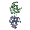 2qfkS S: Starting model for refinement |
|---|---|
| Similar structure data |


-Links
 PDBj
PDBj


- Assembly
| Deposited unit | 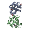
| ||||||||
|---|---|---|---|---|---|---|---|---|---|
| 1 |
| ||||||||
| Unit cell |
|
-Components
| #1: Protein | Mass: 15766.873 Da / Num. of mol.: 2 / Mutation: V59W Source method: isolated from a genetically manipulated source Source: (gene. exp.) Amphitrite ornata (invertebrata) / Production host:  #2: Chemical |   Mass: 616.487 Da / Num. of mol.: 2 / Source method: obtained synthetically / Formula: C34H32FeN4O4 Mass: 616.487 Da / Num. of mol.: 2 / Source method: obtained synthetically / Formula: C34H32FeN4O4#3: Chemical |   Mass: 96.063 Da / Num. of mol.: 2 / Source method: obtained synthetically / Formula: SO4 Mass: 96.063 Da / Num. of mol.: 2 / Source method: obtained synthetically / Formula: SO4#4: Water | ChemComp-HOH / |  Mass: 18.015 Da / Num. of mol.: 342 / Source method: isolated from a natural source / Formula: H2O Mass: 18.015 Da / Num. of mol.: 342 / Source method: isolated from a natural source / Formula: H2OHas ligand of interest | N | |
|---|
-Experimental details
-Experiment
| Experiment | Method: X-RAY DIFFRACTION / Number of used crystals: 1 |
|---|
- Sample preparation
| Crystal | Density Matthews: 2.13 Å3/Da / Density % sol: 42.31 % |
|---|---|
| Crystal grow | Temperature: 277 K / Method: vapor diffusion, hanging drop Details: Protein solution: 8mg/mL protein 20 mM Sodium Cacodylate, pH 6.5 Crystallization: 0.2 M Ammonium Sulfate 30-36% PEG 4000 |
-Data collection
| Diffraction | Mean temperature: 100 K / Serial crystal experiment: N |
|---|---|
| Diffraction source | Source: SYNCHROTRON / Site: APS  / Beamline: 22-BM / Wavelength: 1 Å / Beamline: 22-BM / Wavelength: 1 Å |
| Detector | Type: RAYONIX MX-225 / Detector: CCD / Date: Oct 1, 2010 |
| Radiation | Protocol: SINGLE WAVELENGTH / Monochromatic (M) / Laue (L): M / Scattering type: x-ray |
| Radiation wavelength | Wavelength: 1 Å / Relative weight: 1 |
| Reflection | Resolution: 1.24→48.26 Å / Num. obs: 75146 / % possible obs: 97.5 % / Redundancy: 4.5 % / Rmerge(I) obs: 0.084 / Net I/σ(I): 3.34 |
| Reflection shell | Resolution: 1.24→1.28 Å / Rmerge(I) obs: 0.55 / Num. unique obs: 7502 |
- Processing
| Software |
| ||||||||||||||||||||||||
|---|---|---|---|---|---|---|---|---|---|---|---|---|---|---|---|---|---|---|---|---|---|---|---|---|---|
| Refinement | Method to determine structure: MOLECULAR REPLACEMENT Starting model: 2QFK Resolution: 1.24→48.26 Å / Cor.coef. Fo:Fc: 0.963 / Cor.coef. Fo:Fc free: 0.957 / SU ML: 0 / SU R Cruickshank DPI: 0.0454 / Cross valid method: THROUGHOUT / σ(F): 0 / ESU R: 0.043 / ESU R Free: 0.048 / Stereochemistry target values: MAXIMUM LIKELIHOOD Details: HYDROGENS HAVE BEEN ADDED IN THE RIDING POSITIONS U VALUES : REFINED INDIVIDUALLY
| ||||||||||||||||||||||||
| Solvent computation | Ion probe radii: 0.8 Å / Shrinkage radii: 0.8 Å / VDW probe radii: 1.2 Å / Solvent model: MASK | ||||||||||||||||||||||||
| Displacement parameters | Biso max: 44.6 Å2 / Biso mean: 10.547 Å2 / Biso min: 2.89 Å2
| ||||||||||||||||||||||||
| Refinement step | Cycle: final / Resolution: 1.24→48.26 Å
| ||||||||||||||||||||||||
| LS refinement shell | Resolution: 1.24→1.272 Å / Rfactor Rfree error: 0 / Total num. of bins used: 20
|
