 Movie
Movie Controller

[English] 日本語
 Yorodumi
- PDB-7mlv: Cryo-EM reveals partially and fully assembled native glycine rece... -
+ Open data

- Basic information
| Entry | Database: PDB / ID: 7mlv | ||||||
|---|---|---|---|---|---|---|---|
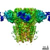
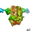
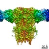
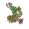
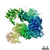
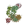
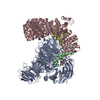
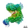
| Title | Cryo-EM reveals partially and fully assembled native glycine receptors,homomeric tetramer | ||||||
 Components Components |
| ||||||
 Keywords Keywords | MEMBRANE PROTEIN / SIGNALING PROTEIN / glycine receptor / ion channel / homomeric tetramer | ||||||
| Function / homology |  Function and homology information Function and homology informationtaurine binding / positive regulation of acrosome reaction / acrosome reaction / negative regulation of transmission of nerve impulse / synaptic transmission, glycinergic / righting reflex / neuromuscular process controlling posture / regulation of respiratory gaseous exchange by nervous system process / extracellularly glycine-gated chloride channel activity / inhibitory synapse ...taurine binding / positive regulation of acrosome reaction / acrosome reaction / negative regulation of transmission of nerve impulse / synaptic transmission, glycinergic / righting reflex / neuromuscular process controlling posture / regulation of respiratory gaseous exchange by nervous system process / extracellularly glycine-gated chloride channel activity / inhibitory synapse / adult walking behavior / glycinergic synapse / excitatory extracellular ligand-gated monoatomic ion channel activity / inhibitory postsynaptic potential / glycine binding / startle response / cellular response to zinc ion / cellular response to ethanol / chloride channel complex / neuronal action potential / muscle contraction / visual perception / ligand-gated monoatomic ion channel activity involved in regulation of presynaptic membrane potential / chloride transmembrane transport / cellular response to amino acid stimulus / transmitter-gated monoatomic ion channel activity involved in regulation of postsynaptic membrane potential / neuropeptide signaling pathway / transmembrane signaling receptor activity / perikaryon / postsynaptic membrane / external side of plasma membrane / dendrite / zinc ion binding Similarity search - Function | ||||||
| Biological species |  | ||||||
| Method | ELECTRON MICROSCOPY / single particle reconstruction / cryo EM / Resolution: 4.1 Å | ||||||
 Authors Authors | Zhu, H. / Gouaux, E. | ||||||
| Funding support |  United States, 1items United States, 1items
| ||||||
 Citation Citation | Journal: Nature / Year: 2021 Title: Architecture and assembly mechanism of native glycine receptors. Authors: Hongtao Zhu / Eric Gouaux / Abstract: Glycine receptors (GlyRs) are pentameric, 'Cys-loop' receptors that form chloride-permeable channels and mediate fast inhibitory signalling throughout the central nervous system. In the spinal cord ...Glycine receptors (GlyRs) are pentameric, 'Cys-loop' receptors that form chloride-permeable channels and mediate fast inhibitory signalling throughout the central nervous system. In the spinal cord and brainstem, GlyRs regulate locomotion and cause movement disorders when mutated. However, the stoichiometry of native GlyRs and the mechanism by which they are assembled remain unclear, despite extensive investigation. Here we report cryo-electron microscopy structures of native GlyRs from pig spinal cord and brainstem, revealing structural insights into heteromeric receptors and their predominant subunit stoichiometry of 4α:1β. Within the heteromeric pentamer, the β(+)-α(-) interface adopts a structure that is distinct from the α(+)-α(-) and α(+)-β(-) interfaces. Furthermore, the β-subunit contains a unique phenylalanine residue that resides within the pore and disrupts the canonical picrotoxin site. These results explain why inclusion of the β-subunit breaks receptor symmetry and alters ion channel pharmacology. We also find incomplete receptor complexes and, by elucidating their structures, reveal the architectures of partially assembled α-trimers and α-tetramers. | ||||||
| History |
|
- Structure visualization
| Movie |
Movie viewer |
|---|---|
| Structure viewer | Molecule: MolmilJmol/JSmol |
- Downloads & links
-Download
| PDBx/mmCIF format | 7mlv.cif.gz | 348.5 KB | Display | PDBx/mmCIF format |
|---|---|---|---|---|
| PDB format | pdb7mlv.ent.gz | 279.2 KB | Display | PDB format |
| PDBx/mmJSON format | 7mlv.json.gz | Tree view | PDBx/mmJSON format | |
| Others |  Other downloads Other downloads |
-Validation report
| Arichive directory | https://data.pdbj.org/pub/pdb/validation_reports/ml/7mlvftp://data.pdbj.org/pub/pdb/validation_reports/ml/7mlv | HTTPS FTP |
|---|
-Related structure data
| Related structure data | 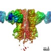 23911MC  7mluC  7mlyC M: map data used to model this data C: citing same article ( |
|---|---|
| Similar structure data |


-Links
 PDBj
PDBj


- Assembly
| Deposited unit | 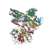
|
|---|---|
| 1 |
|
-Components
-Protein , 1 types, 4 molecules DABC
| #3: Protein | Mass: 52644.750 Da / Num. of mol.: 4 / Source method: isolated from a natural source / Source: (natural) |
|---|
-Antibody , 2 types, 8 molecules JKMIGFLH
| #1: Antibody | Mass: 11743.124 Da / Num. of mol.: 4 Source method: isolated from a genetically manipulated source Source: (gene. exp.) #2: Antibody | Mass: 12983.512 Da / Num. of mol.: 4 Source method: isolated from a genetically manipulated source Source: (gene. exp.) |
|---|
-Sugars , 6 types, 10 molecules 


| #4: Polysaccharide | alpha-D-mannopyranose-(1-3)-[alpha-D-mannopyranose-(1-6)]beta-D-mannopyranose-(1-4)-2-acetamido-2- ...alpha-D-mannopyranose-(1-3)-[alpha-D-mannopyranose-(1-6)]beta-D-mannopyranose-(1-4)-2-acetamido-2-deoxy-beta-D-glucopyranose-(1-4)-2-acetamido-2-deoxy-beta-D-glucopyranose | ||||
|---|---|---|---|---|---|
| #5: Polysaccharide | alpha-D-mannopyranose-(1-3)-beta-D-mannopyranose-(1-4)-2-acetamido-2-deoxy-beta-D-glucopyranose-(1- ...alpha-D-mannopyranose-(1-3)-beta-D-mannopyranose-(1-4)-2-acetamido-2-deoxy-beta-D-glucopyranose-(1-4)-2-acetamido-2-deoxy-beta-D-glucopyranose | ||||
| #6: Polysaccharide | alpha-D-mannopyranose-(1-2)-alpha-D-mannopyranose-(1-6)-alpha-D-mannopyranose-(1-6)-beta-D- ...alpha-D-mannopyranose-(1-2)-alpha-D-mannopyranose-(1-6)-alpha-D-mannopyranose-(1-6)-beta-D-mannopyranose-(1-4)-2-acetamido-2-deoxy-beta-D-glucopyranose-(1-4)-2-acetamido-2-deoxy-beta-D-glucopyranose Type: oligosaccharide / Mass: 1072.964 Da / Num. of mol.: 1 / Source method: isolated from a natural source | ||||
| #7: Sugar | ChemComp-MAN /  Type: D-saccharide, alpha linking / Mass: 180.156 Da / Num. of mol.: 4 / Source method: isolated from a natural source / Formula: C6H12O6 Type: D-saccharide, alpha linking / Mass: 180.156 Da / Num. of mol.: 4 / Source method: isolated from a natural source / Formula: C6H12O6#8: Sugar | ChemComp-BMA / |  Type: D-saccharide, beta linking / Mass: 180.156 Da / Num. of mol.: 1 / Source method: isolated from a natural source / Formula: C6H12O6 Type: D-saccharide, beta linking / Mass: 180.156 Da / Num. of mol.: 1 / Source method: isolated from a natural source / Formula: C6H12O6#9: Sugar |  Type: D-saccharide, beta linking / Mass: 221.208 Da / Num. of mol.: 2 / Source method: isolated from a natural source / Formula: C8H15NO6 Type: D-saccharide, beta linking / Mass: 221.208 Da / Num. of mol.: 2 / Source method: isolated from a natural source / Formula: C8H15NO6 |
-Details
| Has ligand of interest | N |
|---|---|
| Has protein modification | Y |
-Experimental details
-Experiment
| Experiment | Method: ELECTRON MICROSCOPY |
|---|---|
| EM experiment | Aggregation state: PARTICLE / 3D reconstruction method: single particle reconstruction |
- Sample preparation
| Component |
| ||||||||||||||||||||||||
|---|---|---|---|---|---|---|---|---|---|---|---|---|---|---|---|---|---|---|---|---|---|---|---|---|---|
| Molecular weight | Value: 0.4 MDa / Experimental value: YES | ||||||||||||||||||||||||
| Source (natural) |
| ||||||||||||||||||||||||
| Source (recombinant) | Organism: | ||||||||||||||||||||||||
| Buffer solution | pH: 8 | ||||||||||||||||||||||||
| Specimen | Conc.: 0.05 mg/ml / Embedding applied: NO / Shadowing applied: NO / Staining applied: NO / Vitrification applied: YES | ||||||||||||||||||||||||
| Vitrification | Cryogen name: ETHANE |
- Electron microscopy imaging
| Experimental equipment |  Model: Titan Krios / Image courtesy: FEI Company |
|---|---|
| Microscopy | Model: FEI TITAN KRIOS |
| Electron gun | Electron source:  FIELD EMISSION GUN / Accelerating voltage: 300 kV / Illumination mode: FLOOD BEAM FIELD EMISSION GUN / Accelerating voltage: 300 kV / Illumination mode: FLOOD BEAM |
| Electron lens | Mode: BRIGHT FIELD / Cs: 2.7 mm |
| Image recording | Electron dose: 28.2 e/Å2 / Film or detector model: GATAN K3 BIOQUANTUM (6k x 4k) |
- Processing
| EM software |
| ||||||||||||||||||
|---|---|---|---|---|---|---|---|---|---|---|---|---|---|---|---|---|---|---|---|
| CTF correction | Type: PHASE FLIPPING AND AMPLITUDE CORRECTION | ||||||||||||||||||
| Symmetry | Point symmetry: C1 (asymmetric) | ||||||||||||||||||
| 3D reconstruction | Resolution: 4.1 Å / Resolution method: FSC 0.143 CUT-OFF / Num. of particles: 129772 / Symmetry type: POINT |
