 Movie
Movie Controller

[English] 日本語
 Yorodumi
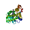
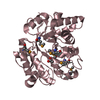
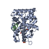
- PDB-7mlo: Crystal structure of ricin A chain in complex with 5-mesitylthiop... -
+ Open data

- Basic information
| Entry | Database: PDB / ID: 7mlo | ||||||
|---|---|---|---|---|---|---|---|
| Title | Crystal structure of ricin A chain in complex with 5-mesitylthiophene-2-carboxylic acid | ||||||
 Components Components | Ricin | ||||||
 Keywords Keywords | Hydrolase/Hydrolase Inhibitor / Ricin chain-A / RTA / Hydrolase / Toxin / Inhibitor / Hydrolase-Hydrolase Inhibitor complex | ||||||
| Function / homology |  Function and homology information Function and homology informationrRNA N-glycosylase / rRNA N-glycosylase activity / AMP binding / defense response / toxin activity / carbohydrate binding / killing of cells of another organism / negative regulation of translation Similarity search - Function | ||||||
| Biological species |  Ricinus communis (castor bean) Ricinus communis (castor bean) | ||||||
| Method |  X-RAY DIFFRACTION / SYNCHROTRON / MOLECULAR REPLACEMENT / Resolution: 1.65 Å X-RAY DIFFRACTION / SYNCHROTRON / MOLECULAR REPLACEMENT / Resolution: 1.65 Å | ||||||
 Authors Authors | Harijan, R.K. / Li, X.P. / Cao, B. / Augeri, D. / Bonanno, J.B. / Almo, S.C. / Tumer, N.E. / Schramm, V.L. | ||||||
| Funding support |  United States, 1items United States, 1items
| ||||||
 Citation Citation | Journal: J.Med.Chem. / Year: 2021 Title: Synthesis and Structural Characterization of Ricin Inhibitors Targeting Ribosome Binding Using Fragment-Based Methods and Structure-Based Design. Authors: Li, X.P. / Harijan, R.K. / Cao, B. / Kahn, J.N. / Pierce, M. / Tsymbal, A.M. / Roberge, J.Y. / Augeri, D. / Tumer, N.E. | ||||||
| History |
|
- Structure visualization
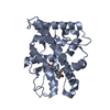
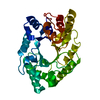
| Structure viewer | Molecule: MolmilJmol/JSmol |
|---|
- Downloads & links
-Download
| PDBx/mmCIF format | 7mlo.cif.gz | 121.9 KB | Display | PDBx/mmCIF format |
|---|---|---|---|---|
| PDB format | pdb7mlo.ent.gz | 92.6 KB | Display | PDB format |
| PDBx/mmJSON format | 7mlo.json.gz | Tree view | PDBx/mmJSON format | |
| Others |  Other downloads Other downloads |
-Validation report
| Arichive directory | https://data.pdbj.org/pub/pdb/validation_reports/ml/7mloftp://data.pdbj.org/pub/pdb/validation_reports/ml/7mlo | HTTPS FTP |
|---|
-Related structure data
| Related structure data |  7mlnC  7mlpC 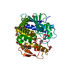 7mltC 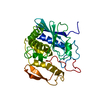 1rtcS S: Starting model for refinement C: citing same article ( |
|---|---|
| Similar structure data |


-Links
 PDBj
PDBj

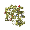
- Assembly
| Deposited unit | 
| ||||||||||||||||||
|---|---|---|---|---|---|---|---|---|---|---|---|---|---|---|---|---|---|---|---|
| 1 | 
| ||||||||||||||||||
| 2 | 
| ||||||||||||||||||
| Unit cell |
| ||||||||||||||||||
| Noncrystallographic symmetry (NCS) | NCS domain:
NCS domain segments: Component-ID: _ / Ens-ID: 1 / Beg auth comp-ID: LYS / Beg label comp-ID: LYS / End auth comp-ID: ALA / End label comp-ID: ALA / Refine code: _ / Auth seq-ID: 4 - 260 / Label seq-ID: 5 - 261
|
-Components
| #1: Protein | Mass: 30067.953 Da / Num. of mol.: 2 Source method: isolated from a genetically manipulated source Source: (gene. exp.) Ricinus communis (castor bean) / Production host:  #2: Chemical | 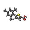  Mass: 246.325 Da / Num. of mol.: 2 / Source method: obtained synthetically / Formula: C14H14O2S / Feature type: SUBJECT OF INVESTIGATION Mass: 246.325 Da / Num. of mol.: 2 / Source method: obtained synthetically / Formula: C14H14O2S / Feature type: SUBJECT OF INVESTIGATION#3: Chemical |   Mass: 62.068 Da / Num. of mol.: 3 / Source method: obtained synthetically / Formula: C2H6O2 Mass: 62.068 Da / Num. of mol.: 3 / Source method: obtained synthetically / Formula: C2H6O2#4: Chemical | ChemComp-SO4 / |   Mass: 96.063 Da / Num. of mol.: 1 / Source method: obtained synthetically / Formula: SO4 Mass: 96.063 Da / Num. of mol.: 1 / Source method: obtained synthetically / Formula: SO4#5: Water | ChemComp-HOH / |  Mass: 18.015 Da / Num. of mol.: 243 / Source method: isolated from a natural source / Formula: H2O Mass: 18.015 Da / Num. of mol.: 243 / Source method: isolated from a natural source / Formula: H2OHas ligand of interest | Y | |
|---|
-Experimental details
-Experiment
| Experiment | Method: X-RAY DIFFRACTION / Number of used crystals: 1 |
|---|
- Sample preparation
| Crystal | Density Matthews: 2.3 Å3/Da / Density % sol: 46.18 % |
|---|---|
| Crystal grow | Temperature: 295 K / Method: vapor diffusion, sitting drop / pH: 6.5 Details: 50 mM Bis-Tris pH 6.5, 50 mM Ammonium Sulfate, 30 %(v/v) Pentaerythritol Ethoxylate (15/4 EO/OH) |
-Data collection
| Diffraction | Mean temperature: 100 K / Serial crystal experiment: N |
|---|---|
| Diffraction source | Source: SYNCHROTRON / Site: APS / Beamline: 31-ID / Wavelength: 0.9793 Å |
| Detector | Type: RAYONIX MX225HE / Detector: CCD / Date: Feb 24, 2021 |
| Radiation | Protocol: SINGLE WAVELENGTH / Monochromatic (M) / Laue (L): M / Scattering type: x-ray |
| Radiation wavelength | Wavelength: 0.9793 Å / Relative weight: 1 |
| Reflection | Resolution: 1.65→126.69 Å / Num. obs: 63861 / % possible obs: 97.7 % / Redundancy: 3.7 % / CC1/2: 0.99 / Rpim(I) all: 0.03 / Net I/σ(I): 11.8 |
| Reflection shell | Resolution: 1.65→1.68 Å / Mean I/σ(I) obs: 1.1 / Num. unique obs: 3060 / CC1/2: 0.55 / Rpim(I) all: 0.59 |
- Processing
| Software |
| ||||||||||||||||||||||||||||||||||||||||||||||||||||||||||||
|---|---|---|---|---|---|---|---|---|---|---|---|---|---|---|---|---|---|---|---|---|---|---|---|---|---|---|---|---|---|---|---|---|---|---|---|---|---|---|---|---|---|---|---|---|---|---|---|---|---|---|---|---|---|---|---|---|---|---|---|---|---|
| Refinement | Method to determine structure: MOLECULAR REPLACEMENT Starting model: 1RTC Resolution: 1.65→126.69 Å / Cor.coef. Fo:Fc: 0.956 / Cor.coef. Fo:Fc free: 0.942 / SU B: 2.923 / SU ML: 0.095 / Cross valid method: THROUGHOUT / σ(F): 0 / ESU R: 0.113 / ESU R Free: 0.108 / Stereochemistry target values: MAXIMUM LIKELIHOOD Details: HYDROGENS HAVE BEEN ADDED IN THE RIDING POSITIONS U VALUES : REFINED INDIVIDUALLY
| ||||||||||||||||||||||||||||||||||||||||||||||||||||||||||||
| Solvent computation | Ion probe radii: 0.8 Å / Shrinkage radii: 0.8 Å / VDW probe radii: 1.2 Å / Solvent model: MASK | ||||||||||||||||||||||||||||||||||||||||||||||||||||||||||||
| Displacement parameters | Biso max: 80.85 Å2 / Biso mean: 29.999 Å2 / Biso min: 15.15 Å2
| ||||||||||||||||||||||||||||||||||||||||||||||||||||||||||||
| Refinement step | Cycle: final / Resolution: 1.65→126.69 Å
| ||||||||||||||||||||||||||||||||||||||||||||||||||||||||||||
| Refine LS restraints |
| ||||||||||||||||||||||||||||||||||||||||||||||||||||||||||||
| Refine LS restraints NCS | Ens-ID: 1 / Number: 8292 / Refine-ID: X-RAY DIFFRACTION / Type: interatomic distance / Rms dev position: 0.09 Å / Weight position: 0.05
| ||||||||||||||||||||||||||||||||||||||||||||||||||||||||||||
| LS refinement shell | Resolution: 1.65→1.693 Å / Rfactor Rfree error: 0 / Total num. of bins used: 20
|
