 Movie
Movie Controller

[English] 日本語
 Yorodumi
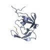
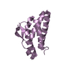
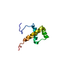
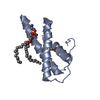
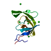
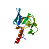
- PDB-7mkk: Crystal structure of Drosophila Panoramix in complex with Sov NTD -
+ Open data

- Basic information
| Entry | Database: PDB / ID: 7mkk | |||||||||
|---|---|---|---|---|---|---|---|---|---|---|
| Title | Crystal structure of Drosophila Panoramix in complex with Sov NTD | |||||||||
 Components Components |
| |||||||||
 Keywords Keywords | RNA BINDING/Metal Binding protein / Piwi / transposon silencing / heterochromatin formation / piRNA pathway / transcriptional silencing / RNA BINDING-Metal Binding protein complex | |||||||||
| Function / homology |  Function and homology information Function and homology informationpiRNA binding / RNA polymerase II transcription repressor complex / transposable element silencing by piRNA-mediated heterochromatin formation / transposable element silencing by heterochromatin formation / transcription repressor complex / heterochromatin formation / positive regulation of transcription by RNA polymerase II / DNA binding / metal ion binding / nucleus Similarity search - Function | |||||||||
| Biological species |  | |||||||||
| Method |  X-RAY DIFFRACTION / SYNCHROTRON / SAD / Resolution: 2.5 Å X-RAY DIFFRACTION / SYNCHROTRON / SAD / Resolution: 2.5 Å | |||||||||
 Authors Authors | Wang, J. / Patel, D.J. | |||||||||
| Funding support |  United States, 2items United States, 2items
| |||||||||
 Citation Citation | Journal: Nat.Struct.Mol.Biol. / Year: 2022 Title: Panoramix SUMOylation on chromatin connects the piRNA pathway to the cellular heterochromatin machinery. Authors: Andreev, V.I. / Yu, C. / Wang, J. / Schnabl, J. / Tirian, L. / Gehre, M. / Handler, D. / Duchek, P. / Novatchkova, M. / Baumgartner, L. / Meixner, K. / Sienski, G. / Patel, D.J. / Brennecke, J. | |||||||||
| History |
|
- Structure visualization
| Structure viewer | Molecule: MolmilJmol/JSmol |
|---|
- Downloads & links
-Download
| PDBx/mmCIF format | 7mkk.cif.gz | 87.1 KB | Display | PDBx/mmCIF format |
|---|---|---|---|---|
| PDB format | pdb7mkk.ent.gz | 67.2 KB | Display | PDB format |
| PDBx/mmJSON format | 7mkk.json.gz | Tree view | PDBx/mmJSON format | |
| Others |  Other downloads Other downloads |
-Validation report
| Arichive directory | https://data.pdbj.org/pub/pdb/validation_reports/mk/7mkkftp://data.pdbj.org/pub/pdb/validation_reports/mk/7mkk | HTTPS FTP |
|---|
-Related structure data
| Similar structure data |
|---|


-Links
 PDBj
PDBj


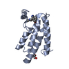
- Assembly
| Deposited unit | 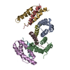
| ||||||||||||
|---|---|---|---|---|---|---|---|---|---|---|---|---|---|
| 1 | 
| ||||||||||||
| 2 | 
| ||||||||||||
| 3 | 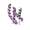
| ||||||||||||
| 4 | 
| ||||||||||||
| Unit cell |
| ||||||||||||
| Components on special symmetry positions |
|
-Components
| #1: Protein | Mass: 9258.927 Da / Num. of mol.: 4 Source method: isolated from a genetically manipulated source Source: (gene. exp.) Gene: sov, Dmel\CG14438, EM25, fs(1)M105, l(1)6Dc, l(1)EA42, l(1)EM25, CG14438, Dmel_CG14438 Production host:  #2: Protein/peptide | Mass: 3094.384 Da / Num. of mol.: 4 Source method: isolated from a genetically manipulated source Source: (gene. exp.) #3: Water | ChemComp-HOH / |  Mass: 18.015 Da / Num. of mol.: 68 / Source method: isolated from a natural source / Formula: H2O Mass: 18.015 Da / Num. of mol.: 68 / Source method: isolated from a natural source / Formula: H2O |
|---|
-Experimental details
-Experiment
| Experiment | Method: X-RAY DIFFRACTION / Number of used crystals: 1 |
|---|
- Sample preparation
| Crystal | Density Matthews: 2.28 Å3/Da / Density % sol: 45.95 % |
|---|---|
| Crystal grow | Temperature: 293 K / Method: vapor diffusion, sitting drop / Details: 0.1 M CHES pH 9.5, 30% (w/v) PEG 3000 |
-Data collection
| Diffraction | Mean temperature: 100 K / Serial crystal experiment: N |
|---|---|
| Diffraction source | Source: SYNCHROTRON / Site: APS / Beamline: 24-ID-E / Wavelength: 0.9792 Å |
| Detector | Type: DECTRIS EIGER X 16M / Detector: PIXEL / Date: Nov 9, 2019 |
| Radiation | Protocol: SINGLE WAVELENGTH / Monochromatic (M) / Laue (L): M / Scattering type: x-ray |
| Radiation wavelength | Wavelength: 0.9792 Å / Relative weight: 1 |
| Reflection | Resolution: 2.5→50 Å / Num. obs: 16430 / % possible obs: 100 % / Redundancy: 12.6 % / CC1/2: 0.999 / Net I/σ(I): 22.8 |
| Reflection shell | Resolution: 2.5→2.64 Å / Num. unique obs: 2329 / CC1/2: 0.93 |
- Processing
| Software |
| |||||||||||||||||||||||||||||||||||
|---|---|---|---|---|---|---|---|---|---|---|---|---|---|---|---|---|---|---|---|---|---|---|---|---|---|---|---|---|---|---|---|---|---|---|---|---|
| Refinement | Method to determine structure: SAD / Resolution: 2.5→47.829 Å / SU ML: 0.37 / Cross valid method: THROUGHOUT / σ(F): 1.35 / Phase error: 27.99 / Stereochemistry target values: ML
| |||||||||||||||||||||||||||||||||||
| Solvent computation | Shrinkage radii: 0.9 Å / VDW probe radii: 1.11 Å / Solvent model: FLAT BULK SOLVENT MODEL | |||||||||||||||||||||||||||||||||||
| Displacement parameters | Biso max: 153.91 Å2 / Biso mean: 67.798 Å2 / Biso min: 26.04 Å2 | |||||||||||||||||||||||||||||||||||
| Refinement step | Cycle: final / Resolution: 2.5→47.829 Å
| |||||||||||||||||||||||||||||||||||
| LS refinement shell | Refine-ID: X-RAY DIFFRACTION / Rfactor Rfree error: 0 / % reflection obs: 100 %
|
