 Movie
Movie Controller

[English] 日本語
 Yorodumi
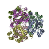
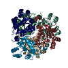
- PDB-7mk0: Trypanosoma cruzi Nucleoside Diphosphate Kinase 1 form a quinary ... -
+ Open data

- Basic information
| Entry | Database: PDB / ID: 7mk0 | |||||||||
|---|---|---|---|---|---|---|---|---|---|---|
| Title | Trypanosoma cruzi Nucleoside Diphosphate Kinase 1 form a quinary multihexameric structure | |||||||||
 Components Components | Nucleoside diphosphate kinase | |||||||||
 Keywords Keywords | TRANSFERASE / Kinase / cruzi / ndpk1 | |||||||||
| Function / homology |  Function and homology information Function and homology informationnucleoside-diphosphate kinase / UTP biosynthetic process / CTP biosynthetic process / nucleoside diphosphate kinase activity / GTP biosynthetic process / ATP binding Similarity search - Function | |||||||||
| Biological species |  | |||||||||
| Method |  X-RAY DIFFRACTION / SYNCHROTRON / MOLECULAR REPLACEMENT / Resolution: 3.5 Å X-RAY DIFFRACTION / SYNCHROTRON / MOLECULAR REPLACEMENT / Resolution: 3.5 Å | |||||||||
 Authors Authors | Gomez, J.A. / Aguilar, C.F. | |||||||||
| Funding support |  Brazil, Brazil,  Argentina, 2items Argentina, 2items
| |||||||||
 Citation Citation | Journal: Acta Crystallogr D Struct Biol / Year: 2022 Title: X-ray diffraction and in vivo studies reveal the quinary structure of Trypanosoma cruzi nucleoside diphosphate kinase 1: a novel helical oligomer structure. Authors: Gomez Barroso, J.A. / Miranda, M.R. / Pereira, C.A. / Garratt, R.C. / Aguilar, C.F. #1: Journal: Acta Crystallographica Section F Structural Biology and Crystallization Communications Year: 2010 Title: Protein preparation, crystallization and preliminary X-ray analysis of Trypanosoma cruzi nucleoside diphosphate kinase 1 Authors: Gomez, J.A. / Aguilar, C.F. | |||||||||
| History |
|
- Structure visualization
| Structure viewer | Molecule: MolmilJmol/JSmol |
|---|
- Downloads & links
-Download
| PDBx/mmCIF format | 7mk0.cif.gz | 656.8 KB | Display | PDBx/mmCIF format |
|---|---|---|---|---|
| PDB format | pdb7mk0.ent.gz | 550.7 KB | Display | PDB format |
| PDBx/mmJSON format | 7mk0.json.gz | Tree view | PDBx/mmJSON format | |
| Others |  Other downloads Other downloads |
-Validation report
| Arichive directory | https://data.pdbj.org/pub/pdb/validation_reports/mk/7mk0ftp://data.pdbj.org/pub/pdb/validation_reports/mk/7mk0 | HTTPS FTP |
|---|
-Related structure data
| Related structure data |  3bbcS S: Starting model for refinement |
|---|---|
| Similar structure data |

-Links
 PDBj
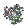
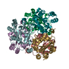
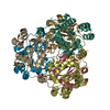
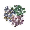
PDBj- Assembly
| Deposited unit | 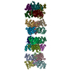
| ||||||||
|---|---|---|---|---|---|---|---|---|---|
| 1 | 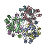
| ||||||||
| 2 | 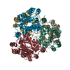
| ||||||||
| 3 | 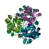
| ||||||||
| 4 | 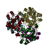
| ||||||||
| Unit cell |
|
-Components
| #1: Protein | Mass: 16742.168 Da / Num. of mol.: 24 Source method: isolated from a genetically manipulated source Source: (gene. exp.)  References: UniProt: A0A2V2WVR0, nucleoside-diphosphate kinase |
|---|
-Experimental details
-Experiment
| Experiment | Method: X-RAY DIFFRACTION / Number of used crystals: 1 |
|---|
- Sample preparation
| Crystal | Density Matthews: 3.23 Å3/Da / Density % sol: 61.98 % |
|---|---|
| Crystal grow | Temperature: 277 K / Method: vapor diffusion, hanging drop / pH: 7.3 / Details: PEG3350 20% Magnesium chloride 200mM |
-Data collection
| Diffraction | Mean temperature: 100 K / Serial crystal experiment: N | |||||||||||||||||||||||||
|---|---|---|---|---|---|---|---|---|---|---|---|---|---|---|---|---|---|---|---|---|---|---|---|---|---|---|
| Diffraction source | Source: SYNCHROTRON / Site: LNLS / Beamline: D03B-MX1 / Wavelength: 1.45 Å | |||||||||||||||||||||||||
| Detector | Type: MAR CCD 165 mm / Detector: CCD / Date: Sep 30, 2000 | |||||||||||||||||||||||||
| Radiation | Protocol: SINGLE WAVELENGTH / Monochromatic (M) / Laue (L): M / Scattering type: x-ray | |||||||||||||||||||||||||
| Radiation wavelength | Wavelength: 1.45 Å / Relative weight: 1 | |||||||||||||||||||||||||
| Reflection twin |
| |||||||||||||||||||||||||
| Reflection | Resolution: 3→86.38 Å / Num. obs: 58612 / % possible obs: 92.3 % / Redundancy: 1.8 % / Rmerge(I) obs: 0.34 / Net I/σ(I): 2.7 | |||||||||||||||||||||||||
| Reflection shell | Resolution: 3.498→3.69 Å / Rmerge(I) obs: 1.121 / Num. unique obs: 9287 / % possible all: 99.9 |
- Processing
| Software |
| ||||||||||||||||||||
|---|---|---|---|---|---|---|---|---|---|---|---|---|---|---|---|---|---|---|---|---|---|
| Refinement | Method to determine structure: MOLECULAR REPLACEMENT Starting model: 3BBC Resolution: 3.5→10 Å / Cor.coef. Fo:Fc: 0.432 / Cor.coef. Fo:Fc free: 0.387 / SU B: 11.722 / SU ML: 0.199 / Cross valid method: THROUGHOUT / σ(F): 0 / ESU R: 0.143 / ESU R Free: 0.148 / Stereochemistry target values: MAXIMUM LIKELIHOOD Details: HYDROGENS HAVE BEEN USED IF PRESENT IN THE INPUT U VALUES : REFINED INDIVIDUALLY
| ||||||||||||||||||||
| Solvent computation | Ion probe radii: 0.8 Å / Shrinkage radii: 0.8 Å / VDW probe radii: 1.2 Å / Solvent model: BABINET MODEL WITH MASK | ||||||||||||||||||||
| Displacement parameters | Biso max: 275.14 Å2 / Biso mean: 15.659 Å2 / Biso min: 2 Å2
| ||||||||||||||||||||
| Refinement step | Cycle: final / Resolution: 3.5→10 Å
| ||||||||||||||||||||
| LS refinement shell | Resolution: 3.5→3.577 Å / Rfactor Rfree error: 0
|
