 Movie
Movie Controller


+ Open data

- Basic information
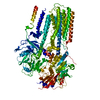
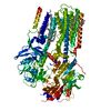
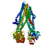
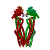
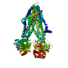
| Entry | Database: PDB / ID: 7mew | ||||||
|---|---|---|---|---|---|---|---|
| Title | E. coli MsbA in complex with G247 | ||||||
 Components Components | ATP-dependent lipid A-core flippase | ||||||
 Keywords Keywords | MEMBRANE PROTEIN | ||||||
| Function / homology |  Function and homology information Function and homology informationABC-type oligopeptide transporter activity / ATPase-coupled lipid transmembrane transporter activity / ATP hydrolysis activity / ATP binding / plasma membrane Similarity search - Function | ||||||
| Biological species |  | ||||||
| Method | ELECTRON MICROSCOPY / single particle reconstruction / cryo EM / Resolution: 3.9 Å | ||||||
 Authors Authors | Thelot, F. / Liao, M. | ||||||
| Funding support |  United States, 1items United States, 1items
| ||||||
 Citation Citation | Journal: Science / Year: 2021 Title: Distinct allosteric mechanisms of first-generation MsbA inhibitors. Authors: François A Thélot / Wenyi Zhang / KangKang Song / Chen Xu / Jing Huang / Maofu Liao /  Abstract: ATP-binding cassette (ABC) transporters couple adenosine 5′-triphosphate (ATP) hydrolysis to substrate transport across biological membranes. Although many are promising drug targets, their ...ATP-binding cassette (ABC) transporters couple adenosine 5′-triphosphate (ATP) hydrolysis to substrate transport across biological membranes. Although many are promising drug targets, their mechanisms of modulation by small-molecule inhibitors remain largely unknown. Two first-generation inhibitors of the MsbA transporter, tetrahydrobenzothiophene 1 (TBT1) and G247, induce opposite effects on ATP hydrolysis. Using single-particle cryo–electron microscopy and functional assays, we show that TBT1 and G247 bind adjacent yet separate pockets in the MsbA transmembrane domains. Two TBT1 molecules asymmetrically occupy the substrate-binding site, which leads to a collapsed inward-facing conformation with decreased distance between the nucleotide-binding domains (NBDs). By contrast, two G247 molecules symmetrically increase NBD distance in a wide inward-open state of MsbA. The divergent mechanisms of action of these MsbA inhibitors provide important insights into ABC transporter pharmacology. | ||||||
| History |
|
- Structure visualization
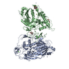
| Movie |
Movie viewer |
|---|---|
| Structure viewer | Molecule: MolmilJmol/JSmol |
- Downloads & links
-Download
| PDBx/mmCIF format | 7mew.cif.gz | 187 KB | Display | PDBx/mmCIF format |
|---|---|---|---|---|
| PDB format | pdb7mew.ent.gz | 139.7 KB | Display | PDB format |
| PDBx/mmJSON format | 7mew.json.gz | Tree view | PDBx/mmJSON format | |
| Others |  Other downloads Other downloads |
-Validation report
| Arichive directory | https://data.pdbj.org/pub/pdb/validation_reports/me/7mewftp://data.pdbj.org/pub/pdb/validation_reports/me/7mew | HTTPS FTP |
|---|
-Related structure data
| Related structure data |  23805MC  7metC  7ritC M: map data used to model this data C: citing same article ( |
|---|---|
| Similar structure data | |
| EM raw data | EMPIAR-10799 (Title: E. coli MsbA in nanodiscs in complex with G247 / Data size: 442.5 Data #1: Motion-corrected micrographs of E. coli MsbA in nanodiscs complex with G247 - Krios Data [micrographs - single frame] Data #2: Motion-corrected micrographs of E. coli MsbA in nanodiscs complex with G247 - Polara Data [micrographs - single frame]) |


-Links
 PDBj
PDBj

- Assembly
| Deposited unit | 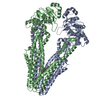
|
|---|---|
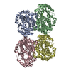
| 1 |
|
-Components
| #1: Protein | Mass: 67310.445 Da / Num. of mol.: 2 Source method: isolated from a genetically manipulated source Source: (gene. exp.) Strain: B / BL21-DE3 / Gene: msbA, ECBD_2681 / Production host: References: UniProt: A0A140NBS6, ABC-type lipid A-core oligosaccharide transporter #2: Chemical | 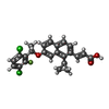  Mass: 445.310 Da / Num. of mol.: 2 / Source method: obtained synthetically / Formula: C24H19Cl2FO3 Mass: 445.310 Da / Num. of mol.: 2 / Source method: obtained synthetically / Formula: C24H19Cl2FO3Has ligand of interest | Y | |
|---|
-Experimental details
-Experiment
| Experiment | Method: ELECTRON MICROSCOPY |
|---|---|
| EM experiment | Aggregation state: PARTICLE / 3D reconstruction method: single particle reconstruction |
- Sample preparation
| Component | Name: E. coli MsbA in complex with G247 / Type: COMPLEX / Entity ID: #1 / Source: RECOMBINANT |
|---|---|
| Molecular weight | Value: 0.13 MDa / Experimental value: NO |
| Source (natural) | Organism: |
| Source (recombinant) | Organism: |
| Buffer solution | pH: 8 |
| Specimen | Embedding applied: NO / Shadowing applied: NO / Staining applied: NO / Vitrification applied: YES |
| Vitrification | Cryogen name: ETHANE |
- Electron microscopy imaging
| Experimental equipment |  Model: Titan Krios / Image courtesy: FEI Company |
|---|---|
| Microscopy | Model: FEI TITAN KRIOS |
| Electron gun | Electron source:  FIELD EMISSION GUN / Accelerating voltage: 300 kV / Illumination mode: OTHER FIELD EMISSION GUN / Accelerating voltage: 300 kV / Illumination mode: OTHER |
| Electron lens | Mode: OTHER |
| Image recording | Electron dose: 63 e/Å2 / Film or detector model: GATAN K2 SUMMIT (4k x 4k) |
- Processing
| CTF correction | Type: PHASE FLIPPING AND AMPLITUDE CORRECTION |
|---|---|
| 3D reconstruction | Resolution: 3.9 Å / Resolution method: FSC 0.143 CUT-OFF / Num. of particles: 349674 / Symmetry type: POINT |
