[English] 日本語
Yorodumi
- PDB-6cxo: Complement component-9 -

+
Open data


ID or keywords:

Loading...

-
Basic information

Entry
Database: PDB / ID: 6cxo
TitleComplement component-9
ComponentsComplement component C9
KeywordsIMMUNE SYSTEM / Immunity / Complex / Complement / MACPF
Function / homology
Function and homology information


Terminal pathway of complement / cell killing / Regulation of Complement cascade / membrane attack complex / cytolysis / other organism cell membrane / complement activation, alternative pathway / complement activation, classical pathway / protein homooligomerization / blood coagulation ...Terminal pathway of complement / cell killing / Regulation of Complement cascade / membrane attack complex / cytolysis / other organism cell membrane / complement activation, alternative pathway / complement activation, classical pathway / protein homooligomerization / blood coagulation / extracellular space / plasma membrane
Similarity search - Function
Complement component C9 / Membrane attack complex component/perforin/complement C9 / Membrane attack complex component/perforin domain, conserved site / Membrane attack complex/perforin (MACPF) domain signature. / membrane-attack complex / perforin / Membrane attack complex/perforin (MACPF) domain profile. / MAC/Perforin domain / Membrane attack complex component/perforin (MACPF) domain / Low-density lipoprotein receptor domain class A / Low-density lipoprotein (LDL) receptor class A, conserved site ...Complement component C9 / Membrane attack complex component/perforin/complement C9 / Membrane attack complex component/perforin domain, conserved site / Membrane attack complex/perforin (MACPF) domain signature. / membrane-attack complex / perforin / Membrane attack complex/perforin (MACPF) domain profile. / MAC/Perforin domain / Membrane attack complex component/perforin (MACPF) domain / Low-density lipoprotein receptor domain class A / Low-density lipoprotein (LDL) receptor class A, conserved site / LDL-receptor class A (LDLRA) domain signature. / LDL-receptor class A (LDLRA) domain profile. / Thrombospondin type 1 domain / Thrombospondin type-1 (TSP1) repeat superfamily / Thrombospondin type-1 (TSP1) repeat profile. / Thrombospondin type 1 repeats / Thrombospondin type-1 (TSP1) repeat / Low-density lipoprotein receptor domain class A / Low-density lipoprotein (LDL) receptor class A repeat / LDL receptor-like superfamily / Growth factor receptor cysteine-rich domain superfamily / EGF-like domain signature 1.
Similarity search - Domain/homology
beta-D-mannopyranose / Complement component C9
Similarity search - Component
Biological speciesMus musculus (house mouse)
MethodX-RAY DIFFRACTION / SYNCHROTRON / MIRAS / Resolution: 2.2 Å
AuthorsLaw, R.H.P. / Spicer, B.A. / Caradoc-Davies, T.T.
Funding support Australia, 1items
OrganizationGrant numberCountry
Australian Research Council (ARC) Australia
Citation
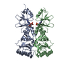
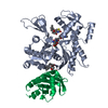
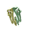
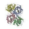
Journal: Nat Commun / Year: 2018
Title: The first transmembrane region of complement component-9 acts as a brake on its self-assembly.
Authors: Bradley A Spicer / Ruby H P Law / Tom T Caradoc-Davies / Sue M Ekkel / Charles Bayly-Jones / Siew-Siew Pang / Paul J Conroy / Georg Ramm / Mazdak Radjainia / Hariprasad Venugopal / James C ...Authors: Bradley A Spicer / Ruby H P Law / Tom T Caradoc-Davies / Sue M Ekkel / Charles Bayly-Jones / Siew-Siew Pang / Paul J Conroy / Georg Ramm / Mazdak Radjainia / Hariprasad Venugopal / James C Whisstock / Michelle A Dunstone /
Abstract: Complement component 9 (C9) functions as the pore-forming component of the Membrane Attack Complex (MAC). During MAC assembly, multiple copies of C9 are sequentially recruited to membrane associated ...Complement component 9 (C9) functions as the pore-forming component of the Membrane Attack Complex (MAC). During MAC assembly, multiple copies of C9 are sequentially recruited to membrane associated C5b8 to form a pore. Here we determined the 2.2 Å crystal structure of monomeric murine C9 and the 3.9 Å resolution cryo EM structure of C9 in a polymeric assembly. Comparison with other MAC proteins reveals that the first transmembrane region (TMH1) in monomeric C9 is uniquely positioned and functions to inhibit its self-assembly in the absence of C5b8. We further show that following C9 recruitment to C5b8, a conformational change in TMH1 permits unidirectional and sequential binding of additional C9 monomers to the growing MAC. This mechanism of pore formation contrasts with related proteins, such as perforin and the cholesterol dependent cytolysins, where it is believed that pre-pore assembly occurs prior to the simultaneous release of the transmembrane regions.
History
DepositionApr 3, 2018Deposition site: RCSB / Processing site: RCSB
Revision 1.0Sep 5, 2018Provider: repository / Type: Initial release
Revision 1.1Jan 1, 2020Group: Author supporting evidence / Category: pdbx_audit_support / Item: _pdbx_audit_support.funding_organization
Revision 1.2Jul 29, 2020Group: Data collection / Derived calculations / Structure summary
Category: chem_comp / entity ...chem_comp / entity / pdbx_chem_comp_identifier / pdbx_entity_nonpoly / pdbx_struct_conn_angle / struct_conn / struct_site / struct_site_gen
Item: _chem_comp.name / _chem_comp.type ..._chem_comp.name / _chem_comp.type / _entity.pdbx_description / _pdbx_entity_nonpoly.name / _pdbx_struct_conn_angle.ptnr1_auth_asym_id / _pdbx_struct_conn_angle.ptnr1_auth_comp_id / _pdbx_struct_conn_angle.ptnr1_auth_seq_id / _pdbx_struct_conn_angle.ptnr1_label_asym_id / _pdbx_struct_conn_angle.ptnr1_label_atom_id / _pdbx_struct_conn_angle.ptnr1_label_comp_id / _pdbx_struct_conn_angle.ptnr1_label_seq_id / _pdbx_struct_conn_angle.ptnr2_auth_comp_id / _pdbx_struct_conn_angle.ptnr2_auth_seq_id / _pdbx_struct_conn_angle.ptnr2_label_asym_id / _pdbx_struct_conn_angle.ptnr2_label_atom_id / _pdbx_struct_conn_angle.ptnr2_label_comp_id / _pdbx_struct_conn_angle.ptnr2_symmetry / _pdbx_struct_conn_angle.ptnr3_auth_asym_id / _pdbx_struct_conn_angle.ptnr3_auth_comp_id / _pdbx_struct_conn_angle.ptnr3_auth_seq_id / _pdbx_struct_conn_angle.ptnr3_label_asym_id / _pdbx_struct_conn_angle.ptnr3_label_atom_id / _pdbx_struct_conn_angle.ptnr3_label_comp_id / _pdbx_struct_conn_angle.ptnr3_label_seq_id / _pdbx_struct_conn_angle.ptnr3_symmetry / _pdbx_struct_conn_angle.value / _struct_conn.conn_type_id / _struct_conn.id / _struct_conn.pdbx_dist_value / _struct_conn.pdbx_leaving_atom_flag / _struct_conn.pdbx_role / _struct_conn.ptnr1_auth_asym_id / _struct_conn.ptnr1_auth_comp_id / _struct_conn.ptnr1_auth_seq_id / _struct_conn.ptnr1_label_asym_id / _struct_conn.ptnr1_label_atom_id / _struct_conn.ptnr1_label_comp_id / _struct_conn.ptnr1_label_seq_id / _struct_conn.ptnr1_symmetry / _struct_conn.ptnr2_auth_asym_id / _struct_conn.ptnr2_auth_comp_id / _struct_conn.ptnr2_auth_seq_id / _struct_conn.ptnr2_label_asym_id / _struct_conn.ptnr2_label_atom_id / _struct_conn.ptnr2_label_comp_id / _struct_conn.ptnr2_symmetry
Description: Carbohydrate remediation / Provider: repository / Type: Remediation

-
Structure visualization

Structure viewerMolecule:
MolmilJmol/JSmol

Downloads & links

-
Assembly

Deposited unit
A: Complement component C9
B: Complement component C9
hetero molecules


Theoretical massNumber of molelcules
Total (without water)121,84615
Polymers120,5882
Non-polymers1,25913
Water6,035335
1
A: Complement component C9
hetero molecules


Theoretical massNumber of molelcules
Total (without water)60,8917
Polymers60,2941
Non-polymers5976
Water181
TypeNameSymmetry operationNumber
identity operation1_555x,y,z1
2
B: Complement component C9
hetero molecules


Theoretical massNumber of molelcules
Total (without water)60,9568
Polymers60,2941
Non-polymers6627
Water181
TypeNameSymmetry operationNumber
identity operation1_555x,y,z1
3
B: Complement component C9
hetero molecules

B: Complement component C9
hetero molecules

A: Complement component C9
hetero molecules

A: Complement component C9
hetero molecules


Theoretical massNumber of molelcules
Total (without water)243,69330
Polymers241,1764
Non-polymers2,51726
Water724
TypeNameSymmetry operationNumber
identity operation1_555x,y,z1
crystal symmetry operation2_756-x+2,y,-z+11
crystal symmetry operation3_575x+1/2,-y+2,-z+1/21
crystal symmetry operation4_675-x+3/2,-y+2,z+1/21
Buried area7470 Å2
ΔGint-335 kcal/mol
Surface area85600 Å2
MethodPISA
Unit cell
Length a, b, c (Å)52.588, 149.213, 165.827
Angle α, β, γ (deg.)90.00, 90.00, 90.00
Int Tables number18
Space group name H-MP21221
Components on special symmetry positions
IDModelComponents
11A-725-

HOH

21A-818-

HOH

-
Components

-
Protein , 1 types, 2 molecules AB

#1: Protein Complement component C9


Mass: 60293.949 Da / Num. of mol.: 2 / Mutation: N28E, N243D, N397D
Source method: isolated from a genetically manipulated source
Source: (gene. exp.) Mus musculus (house mouse) / Gene: C9 / Production host: Homo sapiens (human) / References: UniProt: P06683

-
Sugars , 2 types, 4 molecules

#4: Sugar ChemComp-NAG / 2-acetamido-2-deoxy-beta-D-glucopyranose / N-Acetylglucosamine


Type: D-saccharide, beta linking / Mass: 221.208 Da / Num. of mol.: 2
Source method: isolated from a genetically manipulated source
Formula: C8H15NO6
IdentifierTypeProgram
DGlcpNAcbCONDENSED IUPAC CARBOHYDRATE SYMBOLGMML 1.0
N-acetyl-b-D-glucopyranosamineCOMMON NAMEGMML 1.0
b-D-GlcpNAcIUPAC CARBOHYDRATE SYMBOLPDB-CARE 1.0
GlcNAcSNFG CARBOHYDRATE SYMBOLGMML 1.0
#6: Sugar ChemComp-BMA / beta-D-mannopyranose / Mannose


Type: D-saccharide, beta linking / Mass: 180.156 Da / Num. of mol.: 2
Source method: isolated from a genetically manipulated source
Formula: C6H12O6
IdentifierTypeProgram
DManpbCONDENSED IUPAC CARBOHYDRATE SYMBOLGMML 1.0
b-D-mannopyranoseCOMMON NAMEGMML 1.0
b-D-ManpIUPAC CARBOHYDRATE SYMBOLPDB-CARE 1.0
ManSNFG CARBOHYDRATE SYMBOLGMML 1.0

-
Non-polymers , 4 types, 344 molecules

#2: Chemical
ChemComp-ZN / ZINC ION


Mass: 65.409 Da / Num. of mol.: 5 / Source method: obtained synthetically / Formula: Zn
#3: Chemical ChemComp-CA / CALCIUM ION


Mass: 40.078 Da / Num. of mol.: 2 / Source method: obtained synthetically / Formula: Ca
#5: Chemical ChemComp-MG / MAGNESIUM ION


Mass: 24.305 Da / Num. of mol.: 2 / Source method: obtained synthetically / Formula: Mg
#7: Water ChemComp-HOH / water / Water


Mass: 18.015 Da / Num. of mol.: 335 / Source method: isolated from a natural source / Formula: H2O

-
Experimental details

-
Experiment

ExperimentMethod: X-RAY DIFFRACTION / Number of used crystals: 1

-
Sample preparation

CrystalDensity Matthews: 2.44 Å3/Da / Density % sol: 49.7 % / Description: plate
Crystal growTemperature: 293 K / Method: vapor diffusion, hanging drop / pH: 7.4 / Details: PEG 3350, sodium malonate, zinc chloride / PH range: 7.4

-
Data collection

DiffractionMean temperature: 100 K
Diffraction sourceSource: SYNCHROTRON / Site: Australian Synchrotron / Beamline: MX2 / Wavelength: 1.20365 Å
DetectorType: DECTRIS EIGER X 16M / Detector: PIXEL / Date: Mar 29, 2017
RadiationMonochromator: double crystal / Protocol: SINGLE WAVELENGTH / Monochromatic (M) / Laue (L): M / Scattering type: x-ray
Radiation wavelengthWavelength: 1.20365 Å / Relative weight: 1
ReflectionResolution: 2.2→49.74 Å / Num. obs: 66078 / % possible obs: 98.7 % / Redundancy: 12.1 % / Biso Wilson estimate: 59.38 Å2 / CC1/2: 1 / Rmerge(I) obs: 0.075 / Rpim(I) all: 0.023 / Rrim(I) all: 0.081 / Net I/σ(I): 16.8
Reflection shellResolution: 2.2→2.32 Å / Redundancy: 8.6 % / Rmerge(I) obs: 1.359 / Mean I/σ(I) obs: 1.4 / Num. unique obs: 9323 / CC1/2: 0.656 / Rpim(I) all: 0.514 / % possible all: 97.3

-
Processing

Software
NameVersionClassification
BUSTER2.10.3refinement
XDSdata reduction
XDSdata scaling
CRANK2phasing
RefinementMethod to determine structure: MIRAS / Resolution: 2.2→49.74 Å / Cor.coef. Fo:Fc: 0.942 / Cor.coef. Fo:Fc free: 0.925 / SU R Cruickshank DPI: 0.212 / Cross valid method: THROUGHOUT / σ(F): 0 / SU R Blow DPI: 0.216 / SU Rfree Blow DPI: 0.188 / SU Rfree Cruickshank DPI: 0.187
RfactorNum. reflection% reflectionSelection details
Rfree0.251 3369 5.1 %RANDOM
Rwork0.209 ---
obs0.211 66058 98.2 %-
Displacement parametersBiso mean: 67.54 Å2
Baniso -1Baniso -2Baniso -3
1--8.8409 Å20 Å20 Å2
2---3.2776 Å20 Å2
3---12.1185 Å2
Refine analyzeLuzzati coordinate error obs: 0.32 Å
Refinement stepCycle: 1 / Resolution: 2.2→49.74 Å
ProteinNucleic acidLigandSolventTotal
Num. atoms7160 0 59 335 7554
Refine LS restraints
Refine-IDTypeDev idealNumberRestraint functionWeight
X-RAY DIFFRACTIONt_bond_d0.017390HARMONIC2
X-RAY DIFFRACTIONt_angle_deg0.759999HARMONIC2
X-RAY DIFFRACTIONt_dihedral_angle_d2599SINUSOIDAL2
X-RAY DIFFRACTIONt_incorr_chiral_ct
X-RAY DIFFRACTIONt_pseud_angle
X-RAY DIFFRACTIONt_trig_c_planes198HARMONIC2
X-RAY DIFFRACTIONt_gen_planes1064HARMONIC5
X-RAY DIFFRACTIONt_it7390HARMONIC20
X-RAY DIFFRACTIONt_nbd2SEMIHARMONIC5
X-RAY DIFFRACTIONt_omega_torsion3.05
X-RAY DIFFRACTIONt_other_torsion6.68
X-RAY DIFFRACTIONt_improper_torsion
X-RAY DIFFRACTIONt_chiral_improper_torsion965SEMIHARMONIC5
X-RAY DIFFRACTIONt_sum_occupancies
X-RAY DIFFRACTIONt_utility_distance
X-RAY DIFFRACTIONt_utility_angle
X-RAY DIFFRACTIONt_utility_torsion
X-RAY DIFFRACTIONt_ideal_dist_contact8213SEMIHARMONIC4
LS refinement shellResolution: 2.2→2.26 Å / Total num. of bins used: 20
RfactorNum. reflection% reflection
Rfree0.239 206 4.56 %
Rwork0.226 4314 -
all0.2266 4520 -
obs--92.74 %
Refinement TLS params.

Method: refined / Refine-ID: X-RAY DIFFRACTION

IDL112)L122)L132)L222)L232)L332)S11 (Å °)S12 (Å °)S13 (Å °)S21 (Å °)S22 (Å °)S23 (Å °)S31 (Å °)S32 (Å °)S33 (Å °)T112)T122)T132)T222)T232)T332)Origin x (Å)Origin y (Å)Origin z (Å)
10.5075-0.0391-0.03450.4471-0.22351.62840.0093-0.0926-0.00380.15140.03980.00320.14110.3917-0.0491-0.02170.07120.0305-0.02970.0071-0.12423.841496.028828.0236
20.4577-0.15380.2160.81160.46381.3183-0.0831-0.0002-0.0030.0545-0.07040.10370.0281-0.18440.1535-0.06450.00240.0255-0.0495-0.0117-0.093347.2713133.47755.8762
Refinement TLS group
IDRefine-IDRefine TLS-IDSelection details
1X-RAY DIFFRACTION1{ A|* }
2X-RAY DIFFRACTION2{ B|* }

+
About Yorodumi

-
News

-
Feb 9, 2022. New format data for meta-information of EMDB entries

New format data for meta-information of EMDB entries

  • Version 3 of the EMDB header file is now the official format.
  • The previous official version 1.9 will be removed from the archive.

Related info.:EMDB header

External links:wwPDB to switch to version 3 of the EMDB data model

-
Aug 12, 2020. Covid-19 info

Covid-19 info

URL: https://pdbj.org/emnavi/covid19.php

New page: Covid-19 featured information page in EM Navigator.

Related info.:Covid-19 info / Mar 5, 2020. Novel coronavirus structure data

+
Mar 5, 2020. Novel coronavirus structure data

Novel coronavirus structure data

Related info.:Yorodumi Speices / Aug 12, 2020. Covid-19 info

External links:COVID-19 featured content - PDBj / Molecule of the Month (242):Coronavirus Proteases

+
Jan 31, 2019. EMDB accession codes are about to change! (news from PDBe EMDB page)

EMDB accession codes are about to change! (news from PDBe EMDB page)

  • The allocation of 4 digits for EMDB accession codes will soon come to an end. Whilst these codes will remain in use, new EMDB accession codes will include an additional digit and will expand incrementally as the available range of codes is exhausted. The current 4-digit format prefixed with “EMD-” (i.e. EMD-XXXX) will advance to a 5-digit format (i.e. EMD-XXXXX), and so on. It is currently estimated that the 4-digit codes will be depleted around Spring 2019, at which point the 5-digit format will come into force.
  • The EM Navigator/Yorodumi systems omit the EMD- prefix.

Related info.:Q: What is EMD? / ID/Accession-code notation in Yorodumi/EM Navigator

External links:EMDB Accession Codes are Changing Soon! / Contact to PDBj

+
Jul 12, 2017. Major update of PDB

Major update of PDB

  • wwPDB released updated PDB data conforming to the new PDBx/mmCIF dictionary.
  • This is a major update changing the version number from 4 to 5, and with Remediation, in which all the entries are updated.
  • In this update, many items about electron microscopy experimental information are reorganized (e.g. em_software).
  • Now, EM Navigator and Yorodumi are based on the updated data.

External links:wwPDB Remediation / Enriched Model Files Conforming to OneDep Data Standards Now Available in the PDB FTP Archive

-
Yorodumi

Thousand views of thousand structures

  • Yorodumi is a browser for structure data from EMDB, PDB, SASBDB, etc.
  • This page is also the successor to EM Navigator detail page, and also detail information page/front-end page for Omokage search.
  • The word "yorodu" (or yorozu) is an old Japanese word meaning "ten thousand". "mi" (miru) is to see.

Related info.:EMDB / PDB / SASBDB / Comparison of 3 databanks / Yorodumi Search / Aug 31, 2016. New EM Navigator & Yorodumi / Yorodumi Papers / Jmol/JSmol / Function and homology information / Changes in new EM Navigator and Yorodumi

Read more