Movie
Movie Controller


+ Open data

- Basic information
| Entry | Database: EMDB / ID: EMD-22260 | |||||||||
|---|---|---|---|---|---|---|---|---|---|---|
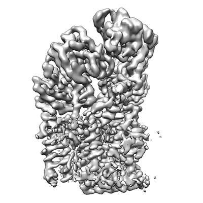
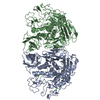
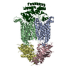
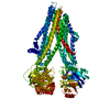
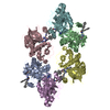
| Title | Structure of P5A-ATPase Spf1, Apo form | |||||||||
 Map data Map data | P5A-ATPase Spf1, Apo form | |||||||||
 Sample Sample |
| |||||||||
 Keywords Keywords | P-type ATPase / transmembrane helix dislocase / protein quality control / endoplasmic reticulum / TRANSPORT PROTEIN | |||||||||
| Function / homology |  Function and homology information Function and homology informationextraction of mislocalized protein from ER membrane / sterol homeostasis / Ion transport by P-type ATPases / membrane protein dislocase activity / intracellular manganese ion homeostasis / P-type ion transporter activity / Translocases; Catalysing the translocation of amino acids and peptides; Linked to the hydrolysis of a nucleoside triphosphate / ATPase-coupled monoatomic cation transmembrane transporter activity / cis-Golgi network / phosphatidylinositol-4-phosphate binding ...extraction of mislocalized protein from ER membrane / sterol homeostasis / Ion transport by P-type ATPases / membrane protein dislocase activity / intracellular manganese ion homeostasis / P-type ion transporter activity / Translocases; Catalysing the translocation of amino acids and peptides; Linked to the hydrolysis of a nucleoside triphosphate / ATPase-coupled monoatomic cation transmembrane transporter activity / cis-Golgi network / phosphatidylinositol-4-phosphate binding / protein hexamerization / protein unfolding / intracellular calcium ion homeostasis / transmembrane transport / protein transport / endoplasmic reticulum membrane / endoplasmic reticulum / ATP hydrolysis activity / mitochondrion / ATP binding / metal ion binding Similarity search - Function | |||||||||
| Biological species |  | |||||||||
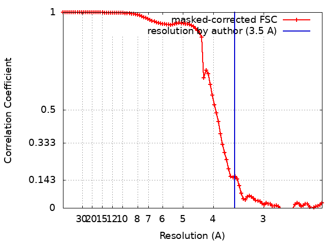
| Method | single particle reconstruction / cryo EM / Resolution: 3.5 Å | |||||||||
 Authors Authors | Park E / Sim SI | |||||||||
| Funding support |  United States, 1 items United States, 1 items
| |||||||||
 Citation Citation | Journal: Science / Year: 2020 Title: The endoplasmic reticulum P5A-ATPase is a transmembrane helix dislocase. Authors: Michael J McKenna / Sue Im Sim / Alban Ordureau / Lianjie Wei / J Wade Harper / Sichen Shao / Eunyong Park / Abstract: Organelle identity depends on protein composition. How mistargeted proteins are selectively recognized and removed from organelles is incompletely understood. Here, we found that the orphan P5A- ...Organelle identity depends on protein composition. How mistargeted proteins are selectively recognized and removed from organelles is incompletely understood. Here, we found that the orphan P5A-adenosine triphosphatase (ATPase) transporter ATP13A1 (Spf1 in yeast) directly interacted with the transmembrane segment (TM) of mitochondrial tail-anchored proteins. P5A-ATPase activity mediated the extraction of mistargeted proteins from the endoplasmic reticulum (ER). Cryo-electron microscopy structures of Spf1 revealed a large, membrane-accessible substrate-binding pocket that alternately faced the ER lumen and cytosol and an endogenous substrate resembling an α-helical TM. Our results indicate that the P5A-ATPase could dislocate misinserted hydrophobic helices flanked by short basic segments from the ER. TM dislocation by the P5A-ATPase establishes an additional class of P-type ATPase substrates and may correct mistakes in protein targeting or topogenesis. | |||||||||
| History |
|
- Structure visualization
| Movie |
Movie viewer |
|---|---|
| Structure viewer | EM map: SurfViewMolmilJmol/JSmol |
| Supplemental images |
- Downloads & links
-EMDB archive
| Map data | emd_22260.map.gz | 32.2 MB | EMDB map data format | |
|---|---|---|---|---|
| Header (meta data) | emd-22260-v30.xmlemd-22260.xml | 23 KB 23 KB | Display Display | EMDB header |
| FSC (resolution estimation) | emd_22260_fsc.xml | 9.2 KB | Display | FSC data file |
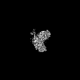
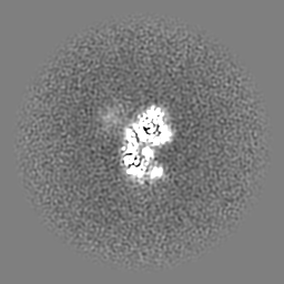
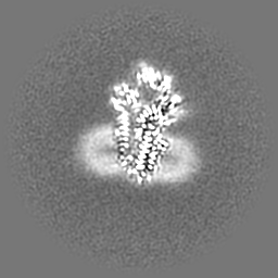
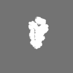
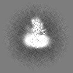
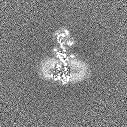
| Images |  emd_22260.png emd_22260.png | 67.2 KB | ||
| Masks | emd_22260_msk_1.map | 64 MB | Mask map | |
| Filedesc metadata | emd-22260.cif.gz | 7 KB | ||
| Others | emd_22260_additional.map.gzemd_22260_half_map_1.map.gzemd_22260_half_map_2.map.gz | 59.7 MB 59.4 MB 59.4 MB | ||
| Archive directory |  http://ftp.pdbj.org/pub/emdb/structures/EMD-22260ftp://ftp.pdbj.org/pub/emdb/structures/EMD-22260 http://ftp.pdbj.org/pub/emdb/structures/EMD-22260ftp://ftp.pdbj.org/pub/emdb/structures/EMD-22260 | HTTPS FTP |
-Related structure data
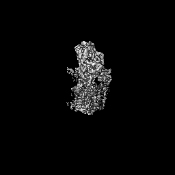
| Related structure data |  6xmpMC  6xmqC  6xmsC  6xmtC  6xmuC M: atomic model generated by this map C: citing same article ( |
|---|---|
| Similar structure data |


-Links
| EMDB pages | EMDB (EBI/PDBe) / EMDataResource |
|---|---|
| Related items in Molecule of the Month |


-Map
| File | Download / File: emd_22260.map.gz / Format: CCP4 / Size: 64 MB / Type: IMAGE STORED AS FLOATING POINT NUMBER (4 BYTES) | ||||||||||||||||||||||||||||||||||||||||||||||||||||||||||||||||||||
|---|---|---|---|---|---|---|---|---|---|---|---|---|---|---|---|---|---|---|---|---|---|---|---|---|---|---|---|---|---|---|---|---|---|---|---|---|---|---|---|---|---|---|---|---|---|---|---|---|---|---|---|---|---|---|---|---|---|---|---|---|---|---|---|---|---|---|---|---|---|
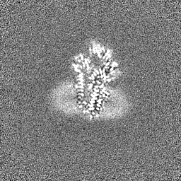
| Annotation | P5A-ATPase Spf1, Apo form | ||||||||||||||||||||||||||||||||||||||||||||||||||||||||||||||||||||
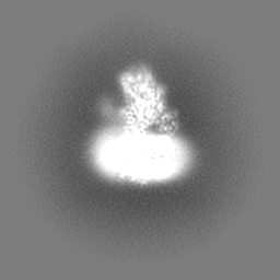
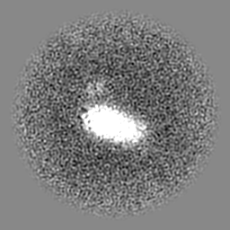
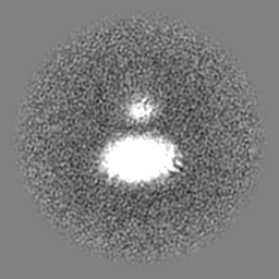
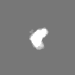
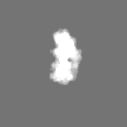
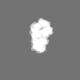
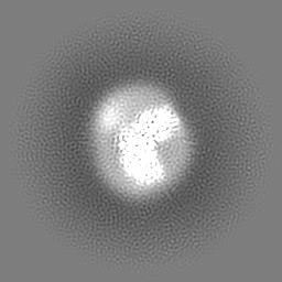
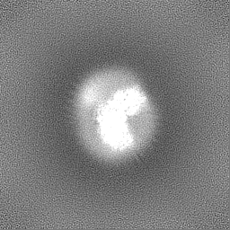
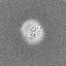
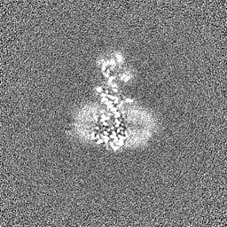
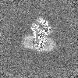
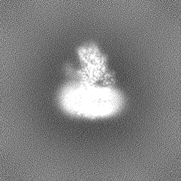
| Projections & slices | Image control
Images are generated by Spider. | ||||||||||||||||||||||||||||||||||||||||||||||||||||||||||||||||||||
| Voxel size | X=Y=Z: 1.14 Å | ||||||||||||||||||||||||||||||||||||||||||||||||||||||||||||||||||||
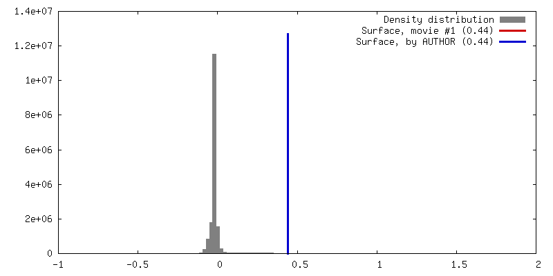
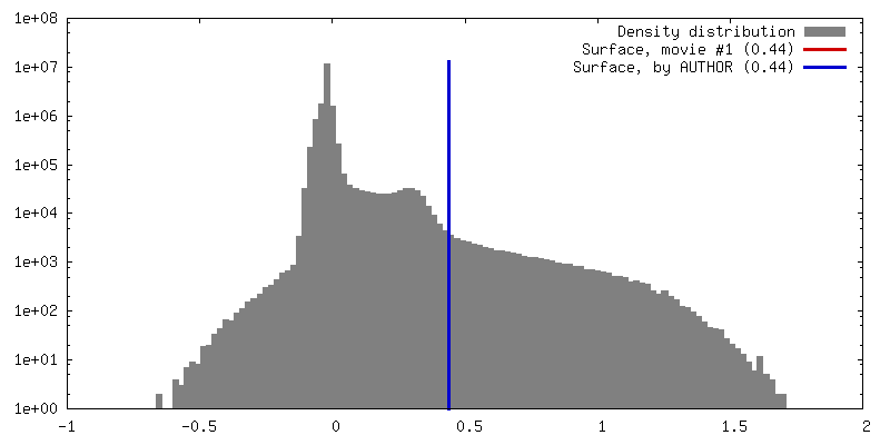
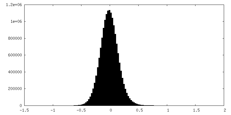
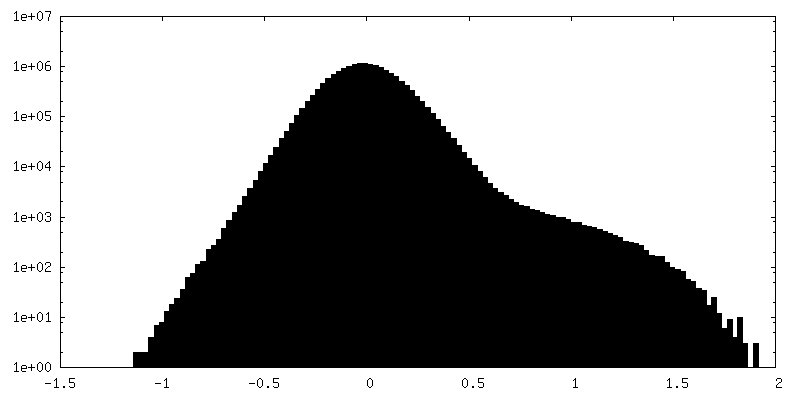
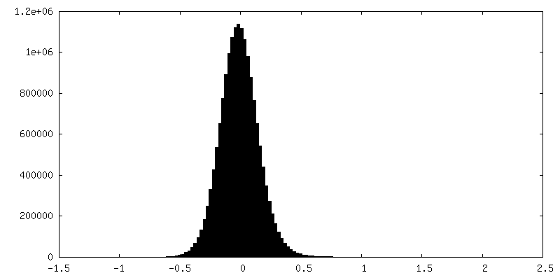
| Density |
| ||||||||||||||||||||||||||||||||||||||||||||||||||||||||||||||||||||
| Symmetry | Space group: 1 | ||||||||||||||||||||||||||||||||||||||||||||||||||||||||||||||||||||
| Details | EMDB XML:
CCP4 map header:
| ||||||||||||||||||||||||||||||||||||||||||||||||||||||||||||||||||||
 Z (Sec.)
 Y (Row.)
 X (Col.)


-Supplemental data
-Mask #1
| File | emd_22260_msk_1.map | ||||||||||||
|---|---|---|---|---|---|---|---|---|---|---|---|---|---|
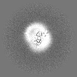
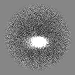
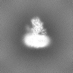
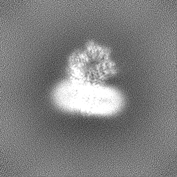
| Projections & Slices |
| ||||||||||||
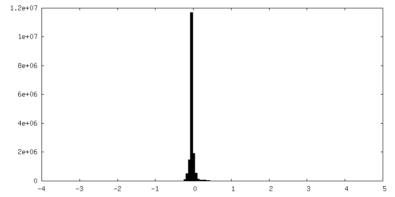
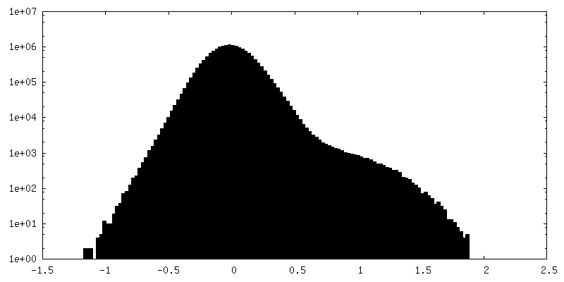
| Density Histograms |


-Additional map: P5A-ATPase Spf1, Apo form
| File | emd_22260_additional.map | ||||||||||||
|---|---|---|---|---|---|---|---|---|---|---|---|---|---|
| Annotation | P5A-ATPase Spf1, Apo form | ||||||||||||
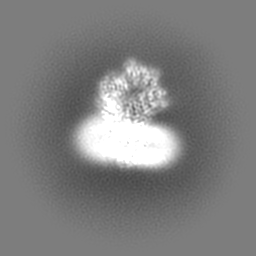
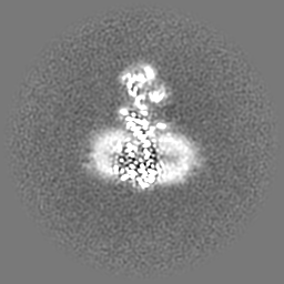
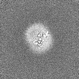
| Projections & Slices |
| ||||||||||||
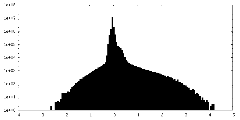
| Density Histograms |

-Half map: P5A-ATPase Spf1, Apo form
| File | emd_22260_half_map_1.map | ||||||||||||
|---|---|---|---|---|---|---|---|---|---|---|---|---|---|
| Annotation | P5A-ATPase Spf1, Apo form | ||||||||||||
| Projections & Slices |
| ||||||||||||
| Density Histograms |

-Half map: P5A-ATPase Spf1, Apo form
| File | emd_22260_half_map_2.map | ||||||||||||
|---|---|---|---|---|---|---|---|---|---|---|---|---|---|
| Annotation | P5A-ATPase Spf1, Apo form | ||||||||||||
| Projections & Slices |
| ||||||||||||
| Density Histograms |

- Sample components
-Entire : P5A-ATPase Spf1 Apo form
| Entire | Name: P5A-ATPase Spf1 Apo form |
|---|---|
| Components |
|
-Supramolecule #1: P5A-ATPase Spf1 Apo form
| Supramolecule | Name: P5A-ATPase Spf1 Apo form / type: complex / ID: 1 / Parent: 0 / Macromolecule list: #1 |
|---|---|
| Source (natural) | Organism: |
-Macromolecule #1: P5A-type ATPase
| Macromolecule | Name: P5A-type ATPase / type: protein_or_peptide / ID: 1 / Number of copies: 1 / Enantiomer: LEVO EC number: Translocases; Catalysing the translocation of inorganic cations; Linked to the hydrolysis of a nucleoside triphosphate |
|---|---|
| Source (natural) | Organism: Strain: ATCC 204508 / S288c |
| Molecular weight | Theoretical: 137.573156 KDa |
| Recombinant expression | Organism: |
| Sequence | String: MTKKSFVSSP IVRDSTLLVP KSLIAKPYVL PFFPLYATFA QLYFQQYDRY IKGPEWTFVY LGTLVSLNIL VMLMPAWNVK IKAKFNYST TKNVNEATHI LIYTTPNNGS DGIVEIQRVT EAGSLQTFFQ FQKKRFLWHE NEQVFSSPKF LVDESPKIGD F QKCKGHSG ...String: MTKKSFVSSP IVRDSTLLVP KSLIAKPYVL PFFPLYATFA QLYFQQYDRY IKGPEWTFVY LGTLVSLNIL VMLMPAWNVK IKAKFNYST TKNVNEATHI LIYTTPNNGS DGIVEIQRVT EAGSLQTFFQ FQKKRFLWHE NEQVFSSPKF LVDESPKIGD F QKCKGHSG DLTHLKRLYG ENSFDIPIPT FMELFKEHAV APLFVFQVFC VALWLLDEFW YYSLFNLFMI ISMEAAAVFQ RL TALKEFR TMGIKPYTIN VFRNKKWVAL QTNELLPMDL VSITRTAEES AIPCDLILLD GSAIVNEAML SGESTPLLKE SIK LRPSED NLQLDGVDKI AVLHGGTKAL QVTPPEHKSD IPPPPDGGAL AIVTKTGFET SQGSLVRVMI YSAERVSVDN KEAL MFILF LLIFAVIASW YVWVEGTKMG RIQSKLILDC ILIITSVVPP ELPMELTMAV NSSLAALAKF YVYCTEPFRI PFAGR IDVC CFDKTGTLTG EDLVFEGLAG ISADSENIRH LYSAAEAPES TILVIGAAHA LVKLEDGDIV GDPMEKATLK AVGWAV ERK NSNYREGTGK LDIIRRFQFS SALKRSASIA SHNDALFAAV KGAPETIRER LSDIPKNYDE IYKSFTRSGS RVLALAS KS LPKMSQSKID DLNRDDVESE LTFNGFLIFH CPLKDDAIET IKMLNESSHR SIMITGDNPL TAVHVAKEVG IVFGETLI L DRAGKSDDNQ LLFRDVEETV SIPFDPSKDT FDHSKLFDRY DIAVTGYALN ALEGHSQLRD LLRHTWVYAR VSPSQKEFL LNTLKDMGYQ TLMCGDGTND VGALKQAHVG IALLNGTEEG LKKLGEQRRL EGMKMMYIKQ TEFMARWNQP QPPVPEPIAH LFPPGPKNP HYLKALESKG TVITPEIRKA VEEANSKPVE VIKPNGLSEK KPADLASLLL NSAGDAQGDE APALKLGDAS C AAPFTSKL ANVSAVTNII RQGRCALVNT IQMYKILALN CLISAYSLSI IYMAGVKFGD GQATVSGLLL SVCFLSISRG KP LEKLSKQ RPQSGIFNVY IMGSILSQFA VHIATLVYIT TEIYKLEPRE PQVDLEKEFA PSLLNTGIFI IQLVQQVSTF AVN YQGEPF RENIRSNKGM YYGLLGVTGL ALASATEFLP ELNEAMKFVP MTDDFKIKLT LTLLLDFFGS WGVEHFFKFF FMDD KPSDI SVQQVKIASK GATGGSTAGG ATTASGTGEN LYFQ UniProtKB: Endoplasmic reticulum transmembrane helix translocase |
-Macromolecule #2: DODECYL-BETA-D-MALTOSIDE
| Macromolecule | Name: DODECYL-BETA-D-MALTOSIDE / type: ligand / ID: 2 / Number of copies: 12 / Formula: LMT |
|---|---|
| Molecular weight | Theoretical: 510.615 Da |
| Chemical component information |  ChemComp-LMT: |
-Experimental details
-Structure determination
| Method | cryo EM |
|---|---|
 Processing Processing | single particle reconstruction |
| Aggregation state | particle |
-Sample preparation
| Concentration | 5 mg/mL |
|---|---|
| Buffer | pH: 7.5 |
| Grid | Model: Quantifoil R1.2/1.3 / Material: GOLD / Mesh: 400 / Pretreatment - Type: GLOW DISCHARGE / Pretreatment - Time: 35 sec. / Pretreatment - Atmosphere: AIR / Pretreatment - Pressure: 0.039 kPa |
| Vitrification | Cryogen name: ETHANE / Chamber humidity: 100 % / Chamber temperature: 277 K |
- Electron microscopy
| Microscope | FEI TALOS ARCTICA |
|---|---|
| Image recording | Film or detector model: GATAN K3 (6k x 4k) / Average electron dose: 50.0 e/Å2 |
| Electron beam | Acceleration voltage: 200 kV / Electron source:  FIELD EMISSION GUN FIELD EMISSION GUN |
| Electron optics | Illumination mode: FLOOD BEAM / Imaging mode: BRIGHT FIELD |
| Experimental equipment |  Model: Talos Arctica / Image courtesy: FEI Company |