Movie
Movie Controller


+ Open data

- Basic information
| Entry | Database: PDB / ID: 7m7b | ||||||||||||
|---|---|---|---|---|---|---|---|---|---|---|---|---|---|
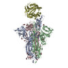
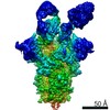
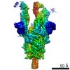
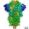
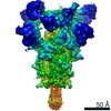
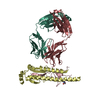
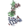
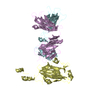
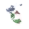
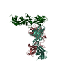
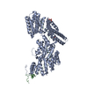
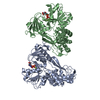
| Title | SARS-CoV-2 Spike:Fab 3D11 complex focused refinement | ||||||||||||
 Components Components |
| ||||||||||||
 Keywords Keywords | VIRAL PROTEIN / SARS-CoV-2 / Spike / antibody / Fab / VIRAL PROTEIN-IMMUNE SYSTEM complex | ||||||||||||
| Function / homology |  Function and homology information Function and homology informationsymbiont-mediated disruption of host tissue / Maturation of spike protein / Translation of Structural Proteins / Virion Assembly and Release / host cell surface / host extracellular region / symbiont-mediated-mediated suppression of host tetherin activity / Induction of Cell-Cell Fusion / structural constituent of virion / positive regulation of viral entry into host cell ...symbiont-mediated disruption of host tissue / Maturation of spike protein / Translation of Structural Proteins / Virion Assembly and Release / host cell surface / host extracellular region / symbiont-mediated-mediated suppression of host tetherin activity / Induction of Cell-Cell Fusion / structural constituent of virion / positive regulation of viral entry into host cell / membrane fusion / host cell endoplasmic reticulum-Golgi intermediate compartment membrane / Attachment and Entry / entry receptor-mediated virion attachment to host cell / receptor-mediated virion attachment to host cell / host cell surface receptor binding / symbiont-mediated suppression of host innate immune response / endocytosis involved in viral entry into host cell / receptor ligand activity / fusion of virus membrane with host plasma membrane / fusion of virus membrane with host endosome membrane / viral envelope / symbiont entry into host cell / virion attachment to host cell / host cell plasma membrane / SARS-CoV-2 activates/modulates innate and adaptive immune responses / virion membrane / membrane / identical protein binding / plasma membrane Similarity search - Function | ||||||||||||
| Biological species |   Severe acute respiratory syndrome coronavirus 2 Severe acute respiratory syndrome coronavirus 2 Homo sapiens (human) Homo sapiens (human) | ||||||||||||
| Method | ELECTRON MICROSCOPY / single particle reconstruction / cryo EM / Resolution: 2.95 Å | ||||||||||||
 Authors Authors | Asarnow, D. / Cheng, Y. | ||||||||||||
| Funding support |  United States, 3items United States, 3items
| ||||||||||||
 Citation Citation | Journal: Cell / Year: 2021 Title: Structural insight into SARS-CoV-2 neutralizing antibodies and modulation of syncytia. Authors: Daniel Asarnow / Bei Wang / Wen-Hsin Lee / Yuanyu Hu / Ching-Wen Huang / Bryan Faust / Patricia Miang Lon Ng / Eve Zi Xian Ngoh / Markus Bohn / David Bulkley / Andrés Pizzorno / Beatrice ...Authors: Daniel Asarnow / Bei Wang / Wen-Hsin Lee / Yuanyu Hu / Ching-Wen Huang / Bryan Faust / Patricia Miang Lon Ng / Eve Zi Xian Ngoh / Markus Bohn / David Bulkley / Andrés Pizzorno / Beatrice Ary / Hwee Ching Tan / Chia Yin Lee / Rabiatul Adawiyah Minhat / Olivier Terrier / Mun Kuen Soh / Frannie Jiuyi Teo / Yvonne Yee Chin Yeap / Shirley Gek Kheng Seah / Conrad En Zuo Chan / Emily Connelly / Nicholas J Young / Sebastian Maurer-Stroh / Laurent Renia / Brendon John Hanson / Manuel Rosa-Calatrava / Aashish Manglik / Yifan Cheng / Charles S Craik / Cheng-I Wang /   Abstract: Infection with severe acute respiratory syndrome coronavirus 2 (SARS-CoV-2) is initiated by binding of the viral Spike protein to host receptor angiotensin-converting enzyme 2 (ACE2), followed by ...Infection with severe acute respiratory syndrome coronavirus 2 (SARS-CoV-2) is initiated by binding of the viral Spike protein to host receptor angiotensin-converting enzyme 2 (ACE2), followed by fusion of viral and host membranes. Although antibodies that block this interaction are in emergency use as early coronavirus disease 2019 (COVID-19) therapies, the precise determinants of neutralization potency remain unknown. We discovered a series of antibodies that potently block ACE2 binding but exhibit divergent neutralization efficacy against the live virus. Strikingly, these neutralizing antibodies can inhibit or enhance Spike-mediated membrane fusion and formation of syncytia, which are associated with chronic tissue damage in individuals with COVID-19. As revealed by cryoelectron microscopy, multiple structures of Spike-antibody complexes have distinct binding modes that not only block ACE2 binding but also alter the Spike protein conformational cycle triggered by ACE2 binding. We show that stabilization of different Spike conformations leads to modulation of Spike-mediated membrane fusion with profound implications for COVID-19 pathology and immunity. | ||||||||||||
| History |
|
- Structure visualization
| Movie |
Movie viewer |
|---|---|
| Structure viewer | Molecule: MolmilJmol/JSmol |
- Downloads & links
-Download
| PDBx/mmCIF format | 7m7b.cif.gz | 163.3 KB | Display | PDBx/mmCIF format |
|---|---|---|---|---|
| PDB format | pdb7m7b.ent.gz | 110.2 KB | Display | PDB format |
| PDBx/mmJSON format | 7m7b.json.gz | Tree view | PDBx/mmJSON format | |
| Others |  Other downloads Other downloads |
-Validation report
| Arichive directory | https://data.pdbj.org/pub/pdb/validation_reports/m7/7m7bftp://data.pdbj.org/pub/pdb/validation_reports/m7/7m7b | HTTPS FTP |
|---|
-Related structure data
| Related structure data |  23709MC  7kqbC  7kqeC  7m71C C: citing same article ( M: map data used to model this data |
|---|---|
| Similar structure data |


-Links
 PDBj
PDBj


- Assembly
| Deposited unit | 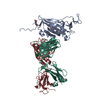
|
|---|---|
| 1 |
|
-Components
| #1: Protein | Mass: 133753.250 Da / Num. of mol.: 1 / Mutation: R682G,R683S,R685S,K986P,V987P Source method: isolated from a genetically manipulated source Source: (gene. exp.) Severe acute respiratory syndrome coronavirus 2Gene: S, 2 / Cell line (production host): ExpiCHO / Production host:   Cricetulus griseus (Chinese hamster) / References: UniProt: P0DTC2 Cricetulus griseus (Chinese hamster) / References: UniProt: P0DTC2 | ||||
|---|---|---|---|---|---|
| #2: Antibody | Mass: 49110.184 Da / Num. of mol.: 1 Source method: isolated from a genetically manipulated source Source: (gene. exp.) Homo sapiens (human) / Cell line (production host): ExpiCHO / Production host: Cricetulus griseus (Chinese hamster) | ||||
| #3: Antibody | Mass: 23055.285 Da / Num. of mol.: 1 Source method: isolated from a genetically manipulated source Source: (gene. exp.) Homo sapiens (human) / Cell line (production host): ExpiCHO / Production host: Cricetulus griseus (Chinese hamster) | ||||
| #4: Sugar |   Type: D-saccharide, beta linking / Mass: 221.208 Da / Num. of mol.: 2 / Source method: obtained synthetically / Formula: C8H15NO6 Type: D-saccharide, beta linking / Mass: 221.208 Da / Num. of mol.: 2 / Source method: obtained synthetically / Formula: C8H15NO6Has ligand of interest | N | Has protein modification | Y | |
-Experimental details
-Experiment
| Experiment | Method: ELECTRON MICROSCOPY |
|---|---|
| EM experiment | Aggregation state: PARTICLE / 3D reconstruction method: single particle reconstruction |
- Sample preparation
| Component |
| ||||||||||||||||||||||||
|---|---|---|---|---|---|---|---|---|---|---|---|---|---|---|---|---|---|---|---|---|---|---|---|---|---|
| Molecular weight | Value: 0.4571 MDa / Experimental value: NO | ||||||||||||||||||||||||
| Source (natural) |
| ||||||||||||||||||||||||
| Source (recombinant) |
| ||||||||||||||||||||||||
| Buffer solution | pH: 8 | ||||||||||||||||||||||||
| Buffer component |
| ||||||||||||||||||||||||
| Specimen | Conc.: 0.4 mg/ml / Embedding applied: NO / Shadowing applied: NO / Staining applied: NO / Vitrification applied: YES | ||||||||||||||||||||||||
| Specimen support | Grid material: GOLD / Grid mesh size: 300 divisions/in. / Grid type: Quantifoil R1.2/1.3 | ||||||||||||||||||||||||
| Vitrification | Instrument: FEI VITROBOT MARK IV / Cryogen name: ETHANE / Humidity: 100 % / Chamber temperature: 4 K / Details: 8-10 seconds, blot force 0 |
- Electron microscopy imaging
| Experimental equipment |  Model: Titan Krios / Image courtesy: FEI Company |
|---|---|
| Microscopy | Model: FEI TITAN KRIOS |
| Electron gun | Electron source:  FIELD EMISSION GUN / Accelerating voltage: 300 kV / Illumination mode: FLOOD BEAM FIELD EMISSION GUN / Accelerating voltage: 300 kV / Illumination mode: FLOOD BEAM |
| Electron lens | Mode: BRIGHT FIELD / Nominal magnification: 105000 X / Nominal defocus max: 1500 nm / Nominal defocus min: 500 nm / Cs: 2.7 mm / C2 aperture diameter: 100 µm / Alignment procedure: ZEMLIN TABLEAU |
| Specimen holder | Cryogen: NITROGEN / Specimen holder model: FEI TITAN KRIOS AUTOGRID HOLDER |
| Image recording | Average exposure time: 5.9 sec. / Electron dose: 67.7 e/Å2 / Film or detector model: GATAN K3 BIOQUANTUM (6k x 4k) / Num. of grids imaged: 1 / Num. of real images: 7536 Details: Movies were collected at super-resolution using correlated double sampling, with a frame time of 0.05 seconds and dose rate 8 e-/px/s |
| EM imaging optics | Energyfilter name: GIF Bioquantum / Energyfilter slit width: 20 eV |
| Image scans | Sampling size: 5 µm / Width: 5760 / Height: 4092 |
- Processing
| EM software |
| ||||||||||||||||||||||||||||||||||||||||
|---|---|---|---|---|---|---|---|---|---|---|---|---|---|---|---|---|---|---|---|---|---|---|---|---|---|---|---|---|---|---|---|---|---|---|---|---|---|---|---|---|---|
| CTF correction | Type: PHASE FLIPPING AND AMPLITUDE CORRECTION | ||||||||||||||||||||||||||||||||||||||||
| Particle selection | Num. of particles selected: 1890759 | ||||||||||||||||||||||||||||||||||||||||
| Symmetry | Point symmetry: C1 (asymmetric) | ||||||||||||||||||||||||||||||||||||||||
| 3D reconstruction | Resolution: 2.95 Å / Resolution method: FSC 0.143 CUT-OFF / Num. of particles: 197150 / Algorithm: FOURIER SPACE / Num. of class averages: 1 / Symmetry type: POINT | ||||||||||||||||||||||||||||||||||||||||
| Atomic model building | Protocol: FLEXIBLE FIT / Space: REAL | ||||||||||||||||||||||||||||||||||||||||
| Atomic model building | PDB-ID: 7A94 Accession code: 7A94 / Source name: PDB / Type: experimental model |