 Movie
Movie Controller

[English] 日本語
 Yorodumi
- PDB-7m75: Room Temperature XFEL Crystallography reveals asymmetry in the vi... -
+ Open data

- Basic information
| Entry | Database: PDB / ID: 7m75 | ||||||||||||||||||||||||
|---|---|---|---|---|---|---|---|---|---|---|---|---|---|---|---|---|---|---|---|---|---|---|---|---|---|
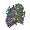
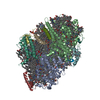
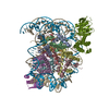
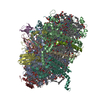
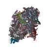
| Title | Room Temperature XFEL Crystallography reveals asymmetry in the vicinity of the two phylloquinones in Photosystem I | ||||||||||||||||||||||||
 Components Components | (Photosystem I ...) x 12 | ||||||||||||||||||||||||
 Keywords Keywords | PHOTOSYNTHESIS / Membrane complex / Electron transport | ||||||||||||||||||||||||
| Function / homology |  Function and homology information Function and homology informationphotosystem I reaction center / photosystem I / photosynthetic electron transport in photosystem I / photosystem I / plasma membrane-derived thylakoid membrane / chlorophyll binding / photosynthesis / 4 iron, 4 sulfur cluster binding / electron transfer activity / oxidoreductase activity ...photosystem I reaction center / photosystem I / photosynthetic electron transport in photosystem I / photosystem I / plasma membrane-derived thylakoid membrane / chlorophyll binding / photosynthesis / 4 iron, 4 sulfur cluster binding / electron transfer activity / oxidoreductase activity / magnesium ion binding / metal ion binding Similarity search - Function | ||||||||||||||||||||||||
| Biological species |   Thermosynechococcus elongatus (bacteria) Thermosynechococcus elongatus (bacteria) | ||||||||||||||||||||||||
| Method |  X-RAY DIFFRACTION / FREE ELECTRON LASER / MOLECULAR REPLACEMENT / Resolution: 2.75 Å X-RAY DIFFRACTION / FREE ELECTRON LASER / MOLECULAR REPLACEMENT / Resolution: 2.75 Å | ||||||||||||||||||||||||
 Authors Authors | Keable, S.M. / Simon, P.S. / Kolsch, A. / Kern, J. / Yachandra, V.K. / Zouni, A. / Yano, J. | ||||||||||||||||||||||||
| Funding support |  United States, United States,  Germany, 7items Germany, 7items
| ||||||||||||||||||||||||
 Citation Citation | Journal: Sci Rep / Year: 2021 Title: Room temperature XFEL crystallography reveals asymmetry in the vicinity of the two phylloquinones in photosystem I. Authors: Keable, S.M. / Kolsch, A. / Simon, P.S. / Dasgupta, M. / Chatterjee, R. / Subramanian, S.K. / Hussein, R. / Ibrahim, M. / Kim, I.S. / Bogacz, I. / Makita, H. / Pham, C.C. / Fuller, F.D. / ...Authors: Keable, S.M. / Kolsch, A. / Simon, P.S. / Dasgupta, M. / Chatterjee, R. / Subramanian, S.K. / Hussein, R. / Ibrahim, M. / Kim, I.S. / Bogacz, I. / Makita, H. / Pham, C.C. / Fuller, F.D. / Gul, S. / Paley, D. / Lassalle, L. / Sutherlin, K.D. / Bhowmick, A. / Moriarty, N.W. / Young, I.D. / Blaschke, J.P. / de Lichtenberg, C. / Chernev, P. / Cheah, M.H. / Park, S. / Park, G. / Kim, J. / Lee, S.J. / Park, J. / Tono, K. / Owada, S. / Hunter, M.S. / Batyuk, A. / Oggenfuss, R. / Sander, M. / Zerdane, S. / Ozerov, D. / Nass, K. / Lemke, H. / Mankowsky, R. / Brewster, A.S. / Messinger, J. / Sauter, N.K. / Yachandra, V.K. / Yano, J. / Zouni, A. / Kern, J. | ||||||||||||||||||||||||
| History |
|
- Structure visualization
| Structure viewer | Molecule: MolmilJmol/JSmol |
|---|
- Downloads & links
-Download
| PDBx/mmCIF format | 7m75.cif.gz | 1.3 MB | Display | PDBx/mmCIF format |
|---|---|---|---|---|
| PDB format | pdb7m75.ent.gz | 1 MB | Display | PDB format |
| PDBx/mmJSON format | 7m75.json.gz | Tree view | PDBx/mmJSON format | |
| Others |  Other downloads Other downloads |
-Validation report
| Arichive directory | https://data.pdbj.org/pub/pdb/validation_reports/m7/7m75ftp://data.pdbj.org/pub/pdb/validation_reports/m7/7m75 | HTTPS FTP |
|---|
-Related structure data
| Related structure data |  7m76C  7m78C  1jb0S S: Starting model for refinement C: citing same article ( |
|---|---|
| Similar structure data |


-Links
 PDBj
PDBj


- Assembly
| Deposited unit | 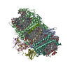
| ||||||||||||
|---|---|---|---|---|---|---|---|---|---|---|---|---|---|
| 1 |
| ||||||||||||
| 2 | 
| ||||||||||||
| Unit cell |
|
-Components
-Photosystem I ... , 12 types, 12 molecules ABCDEFIJKLMX
| #1: Protein | Mass: 83267.773 Da / Num. of mol.: 1 / Source method: isolated from a natural source Source: (natural) Thermosynechococcus elongatus (strain BP-1) (bacteria)Strain: BP-1 / References: UniProt: P0A405, photosystem I |
|---|---|
| #2: Protein | Mass: 82992.453 Da / Num. of mol.: 1 / Source method: isolated from a natural source Source: (natural) Thermosynechococcus elongatus (strain BP-1) (bacteria)Strain: BP-1 / References: UniProt: P0A407, photosystem I |
| #3: Protein | Mass: 8678.011 Da / Num. of mol.: 1 / Source method: isolated from a natural source Source: (natural) Thermosynechococcus elongatus (strain BP-1) (bacteria)Strain: BP-1 / References: UniProt: P0A415, photosystem I |
| #4: Protein | Mass: 15258.297 Da / Num. of mol.: 1 / Source method: isolated from a natural source Source: (natural) Thermosynechococcus elongatus (strain BP-1) (bacteria)Strain: BP-1 / References: UniProt: P0A420 |
| #5: Protein | Mass: 8268.290 Da / Num. of mol.: 1 / Source method: isolated from a natural source Source: (natural) Thermosynechococcus elongatus (strain BP-1) (bacteria)Strain: BP-1 / References: UniProt: P0A423 |
| #6: Protein | Mass: 17716.586 Da / Num. of mol.: 1 / Source method: isolated from a natural source Source: (natural) Thermosynechococcus elongatus (strain BP-1) (bacteria)Strain: BP-1 / References: UniProt: P0A401 |
| #7: Protein/peptide | Mass: 4297.234 Da / Num. of mol.: 1 / Source method: isolated from a natural source Source: (natural) Thermosynechococcus elongatus (strain BP-1) (bacteria)Strain: BP-1 / References: UniProt: P0A427 |
| #8: Protein/peptide | Mass: 4770.698 Da / Num. of mol.: 1 / Source method: isolated from a natural source Source: (natural) Thermosynechococcus elongatus (strain BP-1) (bacteria)Strain: BP-1 / References: UniProt: P0A429 |
| #9: Protein | Mass: 8483.983 Da / Num. of mol.: 1 / Source method: isolated from a natural source Source: (natural) Thermosynechococcus elongatus (strain BP-1) (bacteria)Strain: BP-1 / References: UniProt: P0A425 |
| #10: Protein | Mass: 16156.569 Da / Num. of mol.: 1 / Source method: isolated from a natural source Source: (natural) Thermosynechococcus elongatus (strain BP-1) (bacteria)Strain: BP-1 / References: UniProt: Q8DGB4 |
| #11: Protein/peptide | Mass: 3426.115 Da / Num. of mol.: 1 / Source method: isolated from a natural source Source: (natural) Thermosynechococcus elongatus (strain BP-1) (bacteria)Strain: BP-1 / References: UniProt: P0A403 |
| #12: Protein/peptide | Mass: 3973.744 Da / Num. of mol.: 1 / Source method: isolated from a natural source Source: (natural) Thermosynechococcus elongatus (strain BP-1) (bacteria)Strain: BP-1 / References: UniProt: Q8DKP6 |
-Sugars , 2 types, 2 molecules 

| #21: Sugar | ChemComp-LMT /  Type: D-saccharide / Mass: 510.615 Da / Num. of mol.: 1 / Source method: obtained synthetically / Formula: C24H46O11 / Comment: detergent*YM Type: D-saccharide / Mass: 510.615 Da / Num. of mol.: 1 / Source method: obtained synthetically / Formula: C24H46O11 / Comment: detergent*YM |
|---|---|
| #22: Sugar | ChemComp-DGD /  Type: saccharide / Mass: 949.299 Da / Num. of mol.: 1 / Source method: isolated from a natural source / Formula: C51H96O15 Type: saccharide / Mass: 949.299 Da / Num. of mol.: 1 / Source method: isolated from a natural source / Formula: C51H96O15 |
-Non-polymers , 9 types, 252 molecules 


| #13: Chemical | ChemComp-CL0 /  Mass: 893.489 Da / Num. of mol.: 1 / Source method: obtained synthetically / Formula: C55H72MgN4O5 Mass: 893.489 Da / Num. of mol.: 1 / Source method: obtained synthetically / Formula: C55H72MgN4O5 | ||||||||||||||
|---|---|---|---|---|---|---|---|---|---|---|---|---|---|---|---|
| #14: Chemical | ChemComp-CLA /  Mass: 893.489 Da / Num. of mol.: 95 / Source method: obtained synthetically / Formula: C55H72MgN4O5 Mass: 893.489 Da / Num. of mol.: 95 / Source method: obtained synthetically / Formula: C55H72MgN4O5#15: Chemical |  Mass: 450.696 Da / Num. of mol.: 2 / Source method: obtained synthetically / Formula: C31H46O2 / Feature type: SUBJECT OF INVESTIGATION Mass: 450.696 Da / Num. of mol.: 2 / Source method: obtained synthetically / Formula: C31H46O2 / Feature type: SUBJECT OF INVESTIGATION#16: Chemical | ChemComp-BCR /  Mass: 536.873 Da / Num. of mol.: 22 / Source method: obtained synthetically / Formula: C40H56 Mass: 536.873 Da / Num. of mol.: 22 / Source method: obtained synthetically / Formula: C40H56#17: Chemical | ChemComp-LMG /  Mass: 787.158 Da / Num. of mol.: 4 / Source method: obtained synthetically / Formula: C45H86O10 Mass: 787.158 Da / Num. of mol.: 4 / Source method: obtained synthetically / Formula: C45H86O10#18: Chemical | ChemComp-LHG /  Mass: 722.970 Da / Num. of mol.: 4 / Source method: obtained synthetically / Formula: C38H75O10P / Comment: phospholipid*YM Mass: 722.970 Da / Num. of mol.: 4 / Source method: obtained synthetically / Formula: C38H75O10P / Comment: phospholipid*YM#19: Chemical |  Mass: 351.640 Da / Num. of mol.: 3 / Source method: obtained synthetically / Formula: Fe4S4 Mass: 351.640 Da / Num. of mol.: 3 / Source method: obtained synthetically / Formula: Fe4S4#20: Chemical |  Mass: 40.078 Da / Num. of mol.: 2 / Source method: obtained synthetically / Formula: Ca Mass: 40.078 Da / Num. of mol.: 2 / Source method: obtained synthetically / Formula: Ca#23: Water | ChemComp-HOH / | Mass: 18.015 Da / Num. of mol.: 119 / Source method: isolated from a natural source / Formula: H2O |
-Details
| Has ligand of interest | Y |
|---|---|
| Has protein modification | Y |
-Experimental details
-Experiment
| Experiment | Method: X-RAY DIFFRACTION / Number of used crystals: 1 |
|---|
- Sample preparation
| Crystal | Density Matthews: 7.61 Å3/Da / Density % sol: 83.84 % |
|---|---|
| Crystal grow | Temperature: 298 K / Method: batch mode Details: 5 mM MES, pH 6.0, 12 mM sodium sulfate, 0.2% beta-dodecyl maltoside |
-Data collection
| Diffraction | Mean temperature: 298 K / Serial crystal experiment: N |
|---|---|
| Diffraction source | Source: FREE ELECTRON LASER / Site: SLAC LCLS / Beamline: MFX / Wavelength: 1.305 Å |
| Detector | Type: RAYONIX MX170-HS / Detector: CCD / Date: Jul 3, 2017 |
| Radiation | Protocol: SINGLE WAVELENGTH / Monochromatic (M) / Laue (L): M / Scattering type: x-ray |
| Radiation wavelength | Wavelength: 1.305 Å / Relative weight: 1 |
| Reflection | Resolution: 2.75→56.71 Å / Num. obs: 199647 / % possible obs: 99.98 % / Redundancy: 506.39 % / Biso Wilson estimate: 68.4 Å2 / CC1/2: 0.998 / Net I/σ(I): 3.6 |
| Reflection shell | Resolution: 2.75→2.79 Å / Num. unique obs: 9949 / CC1/2: 0.135 |
- Processing
| Software |
| |||||||||||||||||||||||||||||||||||||||||||||||||||||||||||||||||||||||||||||||||||||||||||||||||||||||||
|---|---|---|---|---|---|---|---|---|---|---|---|---|---|---|---|---|---|---|---|---|---|---|---|---|---|---|---|---|---|---|---|---|---|---|---|---|---|---|---|---|---|---|---|---|---|---|---|---|---|---|---|---|---|---|---|---|---|---|---|---|---|---|---|---|---|---|---|---|---|---|---|---|---|---|---|---|---|---|---|---|---|---|---|---|---|---|---|---|---|---|---|---|---|---|---|---|---|---|---|---|---|---|---|---|---|---|
| Refinement | Method to determine structure: MOLECULAR REPLACEMENT Starting model: PDB entry 1JB0 Resolution: 2.75→56.71 Å / SU ML: 0.5395 / Cross valid method: FREE R-VALUE / σ(F): 1.33 / Phase error: 37.4932 Stereochemistry target values: GeoStd + Monomer Library + CDL v1.2
| |||||||||||||||||||||||||||||||||||||||||||||||||||||||||||||||||||||||||||||||||||||||||||||||||||||||||
| Solvent computation | Shrinkage radii: 0.9 Å / VDW probe radii: 1.11 Å / Solvent model: FLAT BULK SOLVENT MODEL | |||||||||||||||||||||||||||||||||||||||||||||||||||||||||||||||||||||||||||||||||||||||||||||||||||||||||
| Displacement parameters | Biso mean: 77.3 Å2 | |||||||||||||||||||||||||||||||||||||||||||||||||||||||||||||||||||||||||||||||||||||||||||||||||||||||||
| Refinement step | Cycle: LAST / Resolution: 2.75→56.71 Å
| |||||||||||||||||||||||||||||||||||||||||||||||||||||||||||||||||||||||||||||||||||||||||||||||||||||||||
| Refine LS restraints |
| |||||||||||||||||||||||||||||||||||||||||||||||||||||||||||||||||||||||||||||||||||||||||||||||||||||||||
| LS refinement shell |
|
