 Movie
Movie Controller


+ Open data

- Basic information
| Entry | Database: PDB / ID: 7m4u | |||||||||||||||||||||||||||||||||||||||||||||||||||||||||||||||||||||
|---|---|---|---|---|---|---|---|---|---|---|---|---|---|---|---|---|---|---|---|---|---|---|---|---|---|---|---|---|---|---|---|---|---|---|---|---|---|---|---|---|---|---|---|---|---|---|---|---|---|---|---|---|---|---|---|---|---|---|---|---|---|---|---|---|---|---|---|---|---|---|
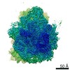
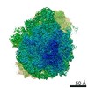
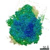
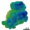
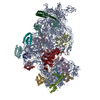
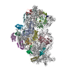
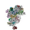
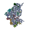
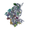
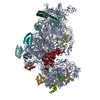
| Title | A. baumannii Ribosome-Eravacycline complex: 30S | |||||||||||||||||||||||||||||||||||||||||||||||||||||||||||||||||||||
 Components Components |
| |||||||||||||||||||||||||||||||||||||||||||||||||||||||||||||||||||||
 Keywords Keywords | RIBOSOME / Acinetobacter baumannii / eravacycline / antibiotic | |||||||||||||||||||||||||||||||||||||||||||||||||||||||||||||||||||||
| Function / homology |  Function and homology information Function and homology informationribosomal small subunit biogenesis / ribosomal small subunit assembly / small ribosomal subunit / small ribosomal subunit rRNA binding / cytosolic small ribosomal subunit / tRNA binding / rRNA binding / structural constituent of ribosome / ribosome / translation ...ribosomal small subunit biogenesis / ribosomal small subunit assembly / small ribosomal subunit / small ribosomal subunit rRNA binding / cytosolic small ribosomal subunit / tRNA binding / rRNA binding / structural constituent of ribosome / ribosome / translation / ribonucleoprotein complex / mRNA binding / RNA binding / cytoplasm / cytosol Similarity search - Function | |||||||||||||||||||||||||||||||||||||||||||||||||||||||||||||||||||||
| Biological species |  Acinetobacter baumannii (bacteria) Acinetobacter baumannii (bacteria) | |||||||||||||||||||||||||||||||||||||||||||||||||||||||||||||||||||||
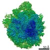
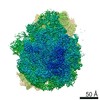
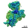
| Method | ELECTRON MICROSCOPY / single particle reconstruction / cryo EM / Resolution: 2.71 Å | |||||||||||||||||||||||||||||||||||||||||||||||||||||||||||||||||||||
 Authors Authors | Morgan, C.E. / Yu, E.W. | |||||||||||||||||||||||||||||||||||||||||||||||||||||||||||||||||||||
| Funding support |  United States, 1items United States, 1items
| |||||||||||||||||||||||||||||||||||||||||||||||||||||||||||||||||||||
 Citation Citation | Journal: mBio / Year: 2021 Title: Cryo-EM Determination of Eravacycline-Bound Structures of the Ribosome and the Multidrug Efflux Pump AdeJ of Acinetobacter baumannii. Authors: Zhemin Zhang / Christopher E Morgan / Robert A Bonomo / Edward W Yu / Abstract: Antibiotic-resistant strains of the Gram-negative pathogen Acinetobacter baumannii have emerged as a significant global health threat. One successful therapeutic option to treat bacterial infections ...Antibiotic-resistant strains of the Gram-negative pathogen Acinetobacter baumannii have emerged as a significant global health threat. One successful therapeutic option to treat bacterial infections has been to target the bacterial ribosome. However, in many cases, multidrug efflux pumps within the bacterium recognize and extrude these clinically important antibiotics designed to inhibit the protein synthesis function of the bacterial ribosome. Thus, multidrug efflux within A. baumannii and other highly drug-resistant strains is a major cause of failure of drug-based treatments of infectious diseases. We here report the first structures of the cinetobacter rug fflux (Ade)J pump in the presence of the antibiotic eravacycline, using single-particle cryo-electron microscopy (cryo-EM). We also describe cryo-EM structures of the eravacycline-bound forms of the A. baumannii ribosome, including the 70S, 50S, and 30S forms. Our data indicate that the AdeJ pump primarily uses hydrophobic interactions to bind eravacycline, while the 70S ribosome utilizes electrostatic interactions to bind this drug. Our work here highlights how an antibiotic can bind multiple bacterial targets through different mechanisms and potentially enables drug optimization by taking advantage of these different modes of ligand binding. Acinetobacter baumannii has developed into a highly antibiotic-resistant Gram-negative pathogen. The prevalent AdeJ multidrug efflux pump mediates resistance to different classes of antibiotics known to inhibit the function of the 70S ribosome. Here, we report the first structures of the A. baumannii AdeJ pump, both in the absence and presence of eravacycline. We also describe structures of the A. baumannii ribosome bound by this antibiotic. Our results indicate that AdeJ and the ribosome use very distinct binding modes for drug recognition. Our work will ultimately enable structure-based drug discovery to combat antibiotic-resistant A. baumannii infection. | |||||||||||||||||||||||||||||||||||||||||||||||||||||||||||||||||||||
| History |
|
- Structure visualization
| Movie |
Movie viewer |
|---|---|
| Structure viewer | Molecule: MolmilJmol/JSmol |
- Downloads & links
-Download
| PDBx/mmCIF format | 7m4u.cif.gz | 1.2 MB | Display | PDBx/mmCIF format |
|---|---|---|---|---|
| PDB format | pdb7m4u.ent.gz | 906.9 KB | Display | PDB format |
| PDBx/mmJSON format | 7m4u.json.gz | Tree view | PDBx/mmJSON format | |
| Others |  Other downloads Other downloads |
-Validation report
| Summary document | 7m4u_validation.pdf.gz | 980.9 KB | Display | wwPDB validaton report |
|---|---|---|---|---|
| Full document | 7m4u_full_validation.pdf.gz | 1 MB | Display | |
| Data in XML | 7m4u_validation.xml.gz | 89.4 KB | Display | |
| Data in CIF | 7m4u_validation.cif.gz | 150.7 KB | Display | |
| Arichive directory | https://data.pdbj.org/pub/pdb/validation_reports/m4/7m4uftp://data.pdbj.org/pub/pdb/validation_reports/m4/7m4u | HTTPS FTP |
-Related structure data
| Related structure data |  23666MC 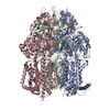 7m4pC  7m4qC 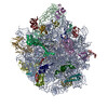 7m4vC 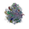 7m4wC 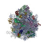 7m4xC 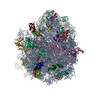 7m4yC 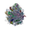 7m4zC M: map data used to model this data C: citing same article ( |
|---|---|
| Similar structure data |


-Links
 PDBj
PDBj


- Assembly
| Deposited unit | 
|
|---|---|
| 1 |
|
-Components
-RNA chain , 1 types, 1 molecules a
| #1: RNA chain | Mass: 500297.531 Da / Num. of mol.: 1 / Source method: isolated from a natural source Source: (natural) Acinetobacter baumannii (strain AB0057) (bacteria)References: GenBank: 1211343212 |
|---|
-30S ribosomal protein ... , 20 types, 20 molecules bcdefghijklmnopqrstu
| #2: Protein | Mass: 27680.357 Da / Num. of mol.: 1 / Source method: isolated from a natural source Source: (natural) Acinetobacter baumannii (strain AB0057) (bacteria)References: UniProt: V5VBC2 |
|---|---|
| #3: Protein | Mass: 27972.461 Da / Num. of mol.: 1 / Source method: isolated from a natural source Source: (natural) Acinetobacter baumannii (strain AB0057) (bacteria)References: UniProt: V5V9N0 |
| #4: Protein | Mass: 23311.818 Da / Num. of mol.: 1 / Source method: isolated from a natural source Source: (natural) Acinetobacter baumannii (strain AB0057) (bacteria)References: UniProt: B7IA15 |
| #5: Protein | Mass: 17181.766 Da / Num. of mol.: 1 / Source method: isolated from a natural source Source: (natural) Acinetobacter baumannii (strain AB0057) (bacteria)References: UniProt: B7IA22 |
| #6: Protein | Mass: 14986.952 Da / Num. of mol.: 1 / Source method: isolated from a natural source Source: (natural) Acinetobacter baumannii (strain AB0057) (bacteria)References: UniProt: B7IBC1 |
| #7: Protein | Mass: 17733.699 Da / Num. of mol.: 1 / Source method: isolated from a natural source Source: (natural) Acinetobacter baumannii (strain AB0057) (bacteria)References: UniProt: B7I7S0 |
| #8: Protein | Mass: 14250.667 Da / Num. of mol.: 1 / Source method: isolated from a natural source Source: (natural) Acinetobacter baumannii (strain AB0057) (bacteria)References: UniProt: B7IA25 |
| #9: Protein | Mass: 14287.610 Da / Num. of mol.: 1 / Source method: isolated from a natural source Source: (natural) Acinetobacter baumannii (strain AB0057) (bacteria)References: UniProt: V5VBA5 |
| #10: Protein | Mass: 11718.531 Da / Num. of mol.: 1 / Source method: isolated from a natural source Source: (natural) Acinetobacter baumannii (strain AB0057) (bacteria)References: UniProt: A0A009L7S8 |
| #11: Protein | Mass: 13558.512 Da / Num. of mol.: 1 / Source method: isolated from a natural source Source: (natural) Acinetobacter baumannii (strain AB0057) (bacteria)References: UniProt: A0A4R0F9S8 |
| #12: Protein | Mass: 13797.134 Da / Num. of mol.: 1 / Source method: isolated from a natural source Source: (natural) Acinetobacter baumannii (strain AB0057) (bacteria)References: UniProt: B7I7R9 |
| #13: Protein | Mass: 13295.635 Da / Num. of mol.: 1 / Source method: isolated from a natural source Source: (natural) Acinetobacter baumannii (strain AB0057) (bacteria)References: UniProt: B7IA17 |
| #14: Protein | Mass: 11438.427 Da / Num. of mol.: 1 / Source method: isolated from a natural source Source: (natural) Acinetobacter baumannii (strain AB0057) (bacteria)References: UniProt: B7IA26 |
| #15: Protein | Mass: 10145.600 Da / Num. of mol.: 1 / Source method: isolated from a natural source Source: (natural) Acinetobacter baumannii (strain AB0057) (bacteria)References: UniProt: B7I3U0 |
| #16: Protein | Mass: 11223.060 Da / Num. of mol.: 1 / Source method: isolated from a natural source Source: (natural) Acinetobacter baumannii (strain AB0057) (bacteria)References: UniProt: A0A1V3DIZ9 |
| #17: Protein | Mass: 9543.101 Da / Num. of mol.: 1 / Source method: isolated from a natural source Source: (natural) Acinetobacter baumannii (strain AB0057) (bacteria)References: UniProt: B7IA30 |
| #18: Protein | Mass: 9009.452 Da / Num. of mol.: 1 / Source method: isolated from a natural source Source: (natural) Acinetobacter baumannii (strain AB0057) (bacteria)References: UniProt: A0A022IPE7 |
| #19: Protein | Mass: 10206.957 Da / Num. of mol.: 1 / Source method: isolated from a natural source Source: (natural) Acinetobacter baumannii (strain AB0057) (bacteria)References: UniProt: B7IA35 |
| #20: Protein | Mass: 9723.420 Da / Num. of mol.: 1 / Source method: isolated from a natural source Source: (natural) Acinetobacter baumannii (strain AB0057) (bacteria)References: UniProt: B7I5N9 |
| #21: Protein | Mass: 8474.033 Da / Num. of mol.: 1 / Source method: isolated from a natural source Source: (natural) Acinetobacter baumannii (strain AB0057) (bacteria)References: UniProt: A0A0Q7FMS9 |
-Non-polymers , 3 types, 275 molecules 


| #22: Chemical | ChemComp-YQM /  Mass: 558.555 Da / Num. of mol.: 1 / Source method: obtained synthetically / Formula: C27H31FN4O8 / Feature type: SUBJECT OF INVESTIGATION Mass: 558.555 Da / Num. of mol.: 1 / Source method: obtained synthetically / Formula: C27H31FN4O8 / Feature type: SUBJECT OF INVESTIGATION | ||
|---|---|---|---|
| #23: Chemical | ChemComp-MG /  Mass: 24.305 Da / Num. of mol.: 83 / Source method: obtained synthetically / Formula: Mg Mass: 24.305 Da / Num. of mol.: 83 / Source method: obtained synthetically / Formula: Mg#24: Water | ChemComp-HOH / | Mass: 18.015 Da / Num. of mol.: 191 / Source method: isolated from a natural source / Formula: H2O |
-Details
| Has ligand of interest | Y |
|---|---|
| Has protein modification | N |
-Experimental details
-Experiment
| Experiment | Method: ELECTRON MICROSCOPY |
|---|---|
| EM experiment | Aggregation state: PARTICLE / 3D reconstruction method: single particle reconstruction |
- Sample preparation
| Component | Name: A baumannii 30S ribosome in complex with Eravacycline / Type: RIBOSOME / Entity ID: #1-#21 / Source: NATURAL |
|---|---|
| Source (natural) | Organism: Acinetobacter baumannii AB0057 (bacteria) |
| Buffer solution | pH: 7.6 |
| Specimen | Embedding applied: NO / Shadowing applied: NO / Staining applied: NO / Vitrification applied: YES |
| Vitrification | Cryogen name: ETHANE |
- Electron microscopy imaging
| Experimental equipment |  Model: Titan Krios / Image courtesy: FEI Company |
|---|---|
| Microscopy | Model: FEI TITAN KRIOS |
| Electron gun | Electron source:  FIELD EMISSION GUN / Accelerating voltage: 300 kV / Illumination mode: SPOT SCAN FIELD EMISSION GUN / Accelerating voltage: 300 kV / Illumination mode: SPOT SCAN |
| Electron lens | Mode: BRIGHT FIELD |
| Image recording | Electron dose: 46 e/Å2 / Film or detector model: GATAN K3 (6k x 4k) |
- Processing
| Software | Name: PHENIX / Version: 1.19_4080: / Classification: refinement | ||||||||||||||||||||||||
|---|---|---|---|---|---|---|---|---|---|---|---|---|---|---|---|---|---|---|---|---|---|---|---|---|---|
| EM software |
| ||||||||||||||||||||||||
| CTF correction | Type: PHASE FLIPPING AND AMPLITUDE CORRECTION | ||||||||||||||||||||||||
| 3D reconstruction | Resolution: 2.71 Å / Resolution method: FSC 0.143 CUT-OFF / Num. of particles: 47367 Details: Resolution of 30S core = 2.80. Resolution of 30S head = 2.71. Symmetry type: POINT | ||||||||||||||||||||||||
| Refine LS restraints |
|
