 Movie
Movie Controller

[English] 日本語
 Yorodumi
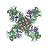
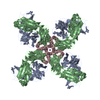
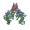
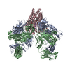
- PDB-7m2h: Structural Snapshots of Intermediates in the Gating of a K+ Channel -
+ Open data

- Basic information
| Entry | Database: PDB / ID: 7m2h | ||||||
|---|---|---|---|---|---|---|---|
| Title | Structural Snapshots of Intermediates in the Gating of a K+ Channel | ||||||
 Components Components |
| ||||||
 Keywords Keywords | TRANSPORT PROTEIN/IMMUNE SYSTEM / Ion channel / K+ channel / pH gated ion channel / TRANSPORT PROTEIN-IMMUNE SYSTEM complex | ||||||
| Function / homology |  Function and homology information Function and homology informationaction potential / voltage-gated potassium channel activity / voltage-gated potassium channel complex / identical protein binding Similarity search - Function | ||||||
| Biological species |   Streptomyces lividans (bacteria) Streptomyces lividans (bacteria) | ||||||
| Method |  X-RAY DIFFRACTION / SYNCHROTRON / MOLECULAR REPLACEMENT / molecular replacement / Resolution: 2.642 Å X-RAY DIFFRACTION / SYNCHROTRON / MOLECULAR REPLACEMENT / molecular replacement / Resolution: 2.642 Å | ||||||
 Authors Authors | Reddi, R. / Valiyaveetil, F.I. | ||||||
| Funding support |  United States, 1items United States, 1items
| ||||||
 Citation Citation | Journal: J.Mol.Biol. / Year: 2021 Title: Structures of Gating Intermediates in a K + channell. Authors: Reddi, R. / Matulef, K. / Riederer, E. / Moenne-Loccoz, P. / Valiyaveetil, F.I. | ||||||
| History |
|
- Structure visualization
| Structure viewer | Molecule: MolmilJmol/JSmol |
|---|
- Downloads & links
-Download
| PDBx/mmCIF format | 7m2h.cif.gz | 219.7 KB | Display | PDBx/mmCIF format |
|---|---|---|---|---|
| PDB format | pdb7m2h.ent.gz | 171.8 KB | Display | PDB format |
| PDBx/mmJSON format | 7m2h.json.gz | Tree view | PDBx/mmJSON format | |
| Others |  Other downloads Other downloads |
-Validation report
| Arichive directory | https://data.pdbj.org/pub/pdb/validation_reports/m2/7m2hftp://data.pdbj.org/pub/pdb/validation_reports/m2/7m2h | HTTPS FTP |
|---|
-Related structure data
| Related structure data | 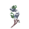 7m2iC 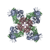 7m2jC  7rp0C  1k4cS S: Starting model for refinement C: citing same article ( |
|---|---|
| Similar structure data |


-Links
 PDBj
PDBj


- Assembly
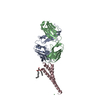
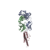
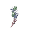
| Deposited unit | 
| |||||||||||||||||||||||||||||||||||||||||||||
|---|---|---|---|---|---|---|---|---|---|---|---|---|---|---|---|---|---|---|---|---|---|---|---|---|---|---|---|---|---|---|---|---|---|---|---|---|---|---|---|---|---|---|---|---|---|---|
| 1 | 
| |||||||||||||||||||||||||||||||||||||||||||||
| 2 | 
| |||||||||||||||||||||||||||||||||||||||||||||
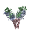
| Unit cell |
| |||||||||||||||||||||||||||||||||||||||||||||
| Components on special symmetry positions |
|
-Components
-Protein , 1 types, 2 molecules CF
| #3: Protein | Mass: 13329.734 Da / Num. of mol.: 2 Source method: isolated from a genetically manipulated source Source: (gene. exp.) Streptomyces lividans (bacteria) / Gene: kcsA, skc1 / Production host: |
|---|
-Antibody , 2 types, 4 molecules ADBE
| #1: Antibody | Mass: 23411.242 Da / Num. of mol.: 2 Source method: isolated from a genetically manipulated source Source: (gene. exp.)  Homo sapiens (human) Homo sapiens (human)#2: Antibody | Mass: 23435.738 Da / Num. of mol.: 2 Source method: isolated from a genetically manipulated source Source: (gene. exp.) Homo sapiens (human) |
|---|
-Non-polymers , 5 types, 145 molecules 


| #4: Chemical |  Mass: 144.254 Da / Num. of mol.: 2 / Source method: obtained synthetically / Formula: C9H20O Mass: 144.254 Da / Num. of mol.: 2 / Source method: obtained synthetically / Formula: C9H20O#5: Chemical | ChemComp-K /  Mass: 39.098 Da / Num. of mol.: 12 / Source method: obtained synthetically / Formula: K / Feature type: SUBJECT OF INVESTIGATION Mass: 39.098 Da / Num. of mol.: 12 / Source method: obtained synthetically / Formula: K / Feature type: SUBJECT OF INVESTIGATION#6: Chemical |  Mass: 625.018 Da / Num. of mol.: 2 / Source method: obtained synthetically / Formula: C39H76O5 Mass: 625.018 Da / Num. of mol.: 2 / Source method: obtained synthetically / Formula: C39H76O5#7: Chemical |  Mass: 242.464 Da / Num. of mol.: 2 / Source method: obtained synthetically / Formula: C16H36N Mass: 242.464 Da / Num. of mol.: 2 / Source method: obtained synthetically / Formula: C16H36N#8: Water | ChemComp-HOH / | Mass: 18.015 Da / Num. of mol.: 127 / Source method: isolated from a natural source / Formula: H2O |
|---|
-Details
| Has ligand of interest | Y |
|---|---|
| Has protein modification | Y |
-Experimental details
-Experiment
| Experiment | Method: X-RAY DIFFRACTION / Number of used crystals: 1 |
|---|
- Sample preparation
| Crystal | Density Matthews: 3.67 Å3/Da / Density % sol: 66.51 % |
|---|---|
| Crystal grow | Temperature: 293 K / Method: vapor diffusion, sitting drop / pH: 6.25 Details: 50 mM MES, pH 6.25, 28% PEG400, 50 mM magnesium acetate PH range: 6.0-6.5 |
-Data collection
| Diffraction | Mean temperature: 100 K / Serial crystal experiment: N |
|---|---|
| Diffraction source | Source: SYNCHROTRON / Site: APS / Beamline: 23-ID-B / Wavelength: 1.04 Å |
| Detector | Type: DECTRIS EIGER X 16M / Detector: PIXEL / Date: Nov 23, 2018 |
| Radiation | Monochromator: Double crystal cryo-cooled Si(111) / Protocol: SINGLE WAVELENGTH / Monochromatic (M) / Laue (L): M / Scattering type: x-ray |
| Radiation wavelength | Wavelength: 1.04 Å / Relative weight: 1 |
| Reflection | Resolution: 2.64→48.801 Å / Num. obs: 54531 / % possible obs: 99.9 % / Redundancy: 6.9 % / CC1/2: 0.99 / Net I/σ(I): 9.1 |
| Reflection shell | Resolution: 2.64→2.73 Å / Mean I/σ(I) obs: 1 / Num. unique obs: 4451 / CC1/2: 0.43 |
-Phasing
| Phasing | Method: molecular replacement |
|---|
- Processing
| Software |
| ||||||||||||||||||||||||||||||||||||||||||||||||||||||||||||||||||||||||||||||||||||
|---|---|---|---|---|---|---|---|---|---|---|---|---|---|---|---|---|---|---|---|---|---|---|---|---|---|---|---|---|---|---|---|---|---|---|---|---|---|---|---|---|---|---|---|---|---|---|---|---|---|---|---|---|---|---|---|---|---|---|---|---|---|---|---|---|---|---|---|---|---|---|---|---|---|---|---|---|---|---|---|---|---|---|---|---|---|
| Refinement | Method to determine structure: MOLECULAR REPLACEMENT Starting model: PDB entry 1K4C Resolution: 2.642→48.801 Å / SU ML: 0.41 / Cross valid method: THROUGHOUT / σ(F): 0 / Phase error: 31.07 / Stereochemistry target values: ML
| ||||||||||||||||||||||||||||||||||||||||||||||||||||||||||||||||||||||||||||||||||||
| Solvent computation | Shrinkage radii: 0.9 Å / VDW probe radii: 1.11 Å / Solvent model: FLAT BULK SOLVENT MODEL | ||||||||||||||||||||||||||||||||||||||||||||||||||||||||||||||||||||||||||||||||||||
| Displacement parameters | Biso max: 106.63 Å2 / Biso mean: 38.3112 Å2 / Biso min: 6.67 Å2 | ||||||||||||||||||||||||||||||||||||||||||||||||||||||||||||||||||||||||||||||||||||
| Refinement step | Cycle: final / Resolution: 2.642→48.801 Å
| ||||||||||||||||||||||||||||||||||||||||||||||||||||||||||||||||||||||||||||||||||||
| LS refinement shell | Refine-ID: X-RAY DIFFRACTION / Rfactor Rfree error: 0
|
