 Movie
Movie Controller

[English] 日本語
 Yorodumi
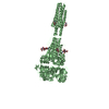
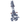
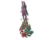
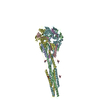
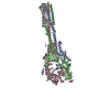
- PDB-7m0i: Crystal structure of a human metapneumovirus subtype B2 trimeric ... -
+ Open data

- Basic information
| Entry | Database: PDB / ID: 7m0i | ||||||
|---|---|---|---|---|---|---|---|
| Title | Crystal structure of a human metapneumovirus subtype B2 trimeric fusion protein | ||||||
 Components Components |
| ||||||
 Keywords Keywords | VIRAL PROTEIN | ||||||
| Function / homology | Precursor fusion glycoprotein F0, Paramyxoviridae / Fusion glycoprotein F0 / fusion of virus membrane with host plasma membrane / host cell plasma membrane / virion membrane / plasma membrane / Fusion glycoprotein F0 Function and homology information Function and homology information | ||||||
| Biological species |  Human metapneumovirus Human metapneumovirus | ||||||
| Method |  X-RAY DIFFRACTION / SYNCHROTRON / MOLECULAR REPLACEMENT / Resolution: 2.81 Å X-RAY DIFFRACTION / SYNCHROTRON / MOLECULAR REPLACEMENT / Resolution: 2.81 Å | ||||||
 Authors Authors | Huang, J. / Mousa, J.J. | ||||||
| Funding support |  United States, 1items United States, 1items
| ||||||
 Citation Citation | Journal: J.Virol. / Year: 2021 Title: Structure, Immunogenicity, and Conformation-Dependent Receptor Binding of the Postfusion Human Metapneumovirus F Protein. Authors: Huang, J. / Chopra, P. / Liu, L. / Nagy, T. / Murray, J. / Tripp, R.A. / Boons, G.J. / Mousa, J.J. | ||||||
| History |
|
- Structure visualization
| Structure viewer | Molecule: MolmilJmol/JSmol |
|---|
- Downloads & links
-Download
| PDBx/mmCIF format | 7m0i.cif.gz | 503.4 KB | Display | PDBx/mmCIF format |
|---|---|---|---|---|
| PDB format | pdb7m0i.ent.gz | 405.5 KB | Display | PDB format |
| PDBx/mmJSON format | 7m0i.json.gz | Tree view | PDBx/mmJSON format | |
| Others |  Other downloads Other downloads |
-Validation report
| Arichive directory | https://data.pdbj.org/pub/pdb/validation_reports/m0/7m0iftp://data.pdbj.org/pub/pdb/validation_reports/m0/7m0i | HTTPS FTP |
|---|
-Related structure data
| Related structure data |  5l1xS S: Starting model for refinement |
|---|---|
| Similar structure data |
-Links
 PDBj
PDBj

- Assembly
| Deposited unit | 
| ||||||||||||
|---|---|---|---|---|---|---|---|---|---|---|---|---|---|
| 1 | 
| ||||||||||||
| 2 | 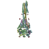
| ||||||||||||
| Unit cell |
|
-Components
| #1: Protein | Mass: 10195.467 Da / Num. of mol.: 6 Source method: isolated from a genetically manipulated source Source: (gene. exp.) Human metapneumovirus / Production host:  Homo sapiens (human) / References: UniProt: C6F474 Homo sapiens (human) / References: UniProt: C6F474#2: Protein | Mass: 46813.914 Da / Num. of mol.: 6 Source method: isolated from a genetically manipulated source Source: (gene. exp.) Human metapneumovirus / Production host: Homo sapiens (human) / References: UniProt: C6F474#3: Sugar | ChemComp-NAG /   Type: D-saccharide, beta linking / Mass: 221.208 Da / Num. of mol.: 18 / Source method: obtained synthetically / Formula: C8H15NO6 Type: D-saccharide, beta linking / Mass: 221.208 Da / Num. of mol.: 18 / Source method: obtained synthetically / Formula: C8H15NO6#4: Water | ChemComp-HOH / |  Mass: 18.015 Da / Num. of mol.: 25 / Source method: isolated from a natural source / Formula: H2O Mass: 18.015 Da / Num. of mol.: 25 / Source method: isolated from a natural source / Formula: H2OHas ligand of interest | N | Has protein modification | Y | Sequence details | hMPV F is a class I fusion glycoprotein, synthesized as an inactive precursor (F0) that needs to be ...hMPV F is a class I fusion glycoprotein, synthesized as an inactive precursor (F0) that needs to be cleaved to become fusion competent. Proteolytic cleavage generates two disulfide-linked subunits (F2 N-terminal and F1 C-terminal fragments) that assemble into a homotrimer. Specifically, REEQIENPRQ | |
|---|
-Experimental details
-Experiment
| Experiment | Method: X-RAY DIFFRACTION / Number of used crystals: 1 |
|---|
- Sample preparation
| Crystal | Density Matthews: 4.63 Å3/Da / Density % sol: 73.46 % |
|---|---|
| Crystal grow | Temperature: 298 K / Method: vapor diffusion, sitting drop / Details: 0.1 M Tris pH 8.5, 2.0 M Ammonium sulfate |
-Data collection
| Diffraction | Mean temperature: 100 K / Serial crystal experiment: N |
|---|---|
| Diffraction source | Source: SYNCHROTRON / Site: APS / Beamline: 21-ID-D / Wavelength: 1 Å |
| Detector | Type: DECTRIS EIGER X 16M / Detector: PIXEL / Date: Oct 23, 2019 |
| Radiation | Protocol: SINGLE WAVELENGTH / Monochromatic (M) / Laue (L): M / Scattering type: x-ray |
| Radiation wavelength | Wavelength: 1 Å / Relative weight: 1 |
| Reflection | Resolution: 2.81→49.69 Å / Num. obs: 155097 / % possible obs: 75.6 % / Redundancy: 14.9 % / Biso Wilson estimate: 34.4 Å2 / CC1/2: 0.973 / CC star: 0.993 / Net I/σ(I): 6.5 |
| Reflection shell | Resolution: 2.811→2.911 Å / Redundancy: 14.5 % / Mean I/σ(I) obs: 0.77 / Num. unique obs: 1294 / CC1/2: 0.227 |
- Processing
| Software |
| |||||||||||||||||||||||||||||||||||||||||||||||||||||||||||||||||||||||||||||||||||||||||||||||||||||||||||||||||||||||||||||||||||||||||||||||||||||||||||||||||||||||||||||||||||||||||||||||||||||||||||||||||||||||||
|---|---|---|---|---|---|---|---|---|---|---|---|---|---|---|---|---|---|---|---|---|---|---|---|---|---|---|---|---|---|---|---|---|---|---|---|---|---|---|---|---|---|---|---|---|---|---|---|---|---|---|---|---|---|---|---|---|---|---|---|---|---|---|---|---|---|---|---|---|---|---|---|---|---|---|---|---|---|---|---|---|---|---|---|---|---|---|---|---|---|---|---|---|---|---|---|---|---|---|---|---|---|---|---|---|---|---|---|---|---|---|---|---|---|---|---|---|---|---|---|---|---|---|---|---|---|---|---|---|---|---|---|---|---|---|---|---|---|---|---|---|---|---|---|---|---|---|---|---|---|---|---|---|---|---|---|---|---|---|---|---|---|---|---|---|---|---|---|---|---|---|---|---|---|---|---|---|---|---|---|---|---|---|---|---|---|---|---|---|---|---|---|---|---|---|---|---|---|---|---|---|---|---|---|---|---|---|---|---|---|---|---|---|---|---|---|---|---|---|
| Refinement | Method to determine structure: MOLECULAR REPLACEMENT Starting model: 5L1X Resolution: 2.81→49.69 Å / SU ML: 0.3906 / Cross valid method: FREE R-VALUE / σ(F): 1.35 / Phase error: 28.9656 Stereochemistry target values: GeoStd + Monomer Library + CDL v1.2
| |||||||||||||||||||||||||||||||||||||||||||||||||||||||||||||||||||||||||||||||||||||||||||||||||||||||||||||||||||||||||||||||||||||||||||||||||||||||||||||||||||||||||||||||||||||||||||||||||||||||||||||||||||||||||
| Solvent computation | Shrinkage radii: 0.9 Å / VDW probe radii: 1.11 Å / Solvent model: FLAT BULK SOLVENT MODEL | |||||||||||||||||||||||||||||||||||||||||||||||||||||||||||||||||||||||||||||||||||||||||||||||||||||||||||||||||||||||||||||||||||||||||||||||||||||||||||||||||||||||||||||||||||||||||||||||||||||||||||||||||||||||||
| Displacement parameters | Biso mean: 39.29 Å2 | |||||||||||||||||||||||||||||||||||||||||||||||||||||||||||||||||||||||||||||||||||||||||||||||||||||||||||||||||||||||||||||||||||||||||||||||||||||||||||||||||||||||||||||||||||||||||||||||||||||||||||||||||||||||||
| Refinement step | Cycle: LAST / Resolution: 2.81→49.69 Å
| |||||||||||||||||||||||||||||||||||||||||||||||||||||||||||||||||||||||||||||||||||||||||||||||||||||||||||||||||||||||||||||||||||||||||||||||||||||||||||||||||||||||||||||||||||||||||||||||||||||||||||||||||||||||||
| Refine LS restraints |
| |||||||||||||||||||||||||||||||||||||||||||||||||||||||||||||||||||||||||||||||||||||||||||||||||||||||||||||||||||||||||||||||||||||||||||||||||||||||||||||||||||||||||||||||||||||||||||||||||||||||||||||||||||||||||
| LS refinement shell |
|
