 Movie
Movie Controller

[English] 日本語
 Yorodumi
- PDB-7lrn: Structure of the Siderophore Interacting Protein from Acinetbacte... -
+ Open data

- Basic information
| Entry | Database: PDB / ID: 7lrn | ||||||
|---|---|---|---|---|---|---|---|
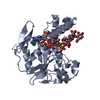
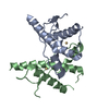
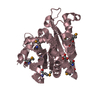
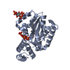
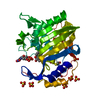
| Title | Structure of the Siderophore Interacting Protein from Acinetbacter baumannii | ||||||
 Components Components | NADPH-dependent ferric siderophore reductase | ||||||
 Keywords Keywords | OXIDOREDUCTASE / SIDEROPHORE-INTERACTING PROTEIN / FLAVIN BINDING | ||||||
| Function / homology |  Function and homology information Function and homology informationferric-chelate reductase (NADPH) activity / cellular response to iron ion starvation / iron import into cell / siderophore transport / FAD binding Similarity search - Function | ||||||
| Biological species |  Acinetobacter baumannii (bacteria) Acinetobacter baumannii (bacteria) | ||||||
| Method |  X-RAY DIFFRACTION / SYNCHROTRON / MOLECULAR REPLACEMENT / Resolution: 2.85 Å X-RAY DIFFRACTION / SYNCHROTRON / MOLECULAR REPLACEMENT / Resolution: 2.85 Å | ||||||
 Authors Authors | Tanner, J.J. / Korasick, D.A. | ||||||
| Funding support |  United States, 1items United States, 1items
| ||||||
 Citation Citation | Journal: Acs Omega / Year: 2021 Title: Structural and Biochemical Characterization of the Flavin-Dependent Siderophore-Interacting Protein from Acinetobacter baumannii . Authors: Valentino, H. / Korasick, D.A. / Bohac, T.J. / Shapiro, J.A. / Wencewicz, T.A. / Tanner, J.J. / Sobrado, P. | ||||||
| History |
|
- Structure visualization
| Structure viewer | Molecule: MolmilJmol/JSmol |
|---|
- Downloads & links
-Download
| PDBx/mmCIF format | 7lrn.cif.gz | 212.6 KB | Display | PDBx/mmCIF format |
|---|---|---|---|---|
| PDB format | pdb7lrn.ent.gz | 167.8 KB | Display | PDB format |
| PDBx/mmJSON format | 7lrn.json.gz | Tree view | PDBx/mmJSON format | |
| Others |  Other downloads Other downloads |
-Validation report
| Arichive directory | https://data.pdbj.org/pub/pdb/validation_reports/lr/7lrnftp://data.pdbj.org/pub/pdb/validation_reports/lr/7lrn | HTTPS FTP |
|---|
-Related structure data
| Related structure data |  2gpjS S: Starting model for refinement |
|---|---|
| Similar structure data |


-Links
 PDBj
PDBj- Assembly
| Deposited unit | 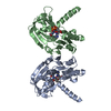
| |||||||||||||||||||||||||||||||||||||||||||||||||||||||||||||||||||||||||||||||||||||||||||||
|---|---|---|---|---|---|---|---|---|---|---|---|---|---|---|---|---|---|---|---|---|---|---|---|---|---|---|---|---|---|---|---|---|---|---|---|---|---|---|---|---|---|---|---|---|---|---|---|---|---|---|---|---|---|---|---|---|---|---|---|---|---|---|---|---|---|---|---|---|---|---|---|---|---|---|---|---|---|---|---|---|---|---|---|---|---|---|---|---|---|---|---|---|---|---|
| 1 | 
| |||||||||||||||||||||||||||||||||||||||||||||||||||||||||||||||||||||||||||||||||||||||||||||
| 2 | 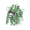
| |||||||||||||||||||||||||||||||||||||||||||||||||||||||||||||||||||||||||||||||||||||||||||||
| Unit cell |
| |||||||||||||||||||||||||||||||||||||||||||||||||||||||||||||||||||||||||||||||||||||||||||||
| Noncrystallographic symmetry (NCS) | NCS domain:
NCS domain segments: Ens-ID: 1
|