 Movie
Movie Controller

[English] 日本語
 Yorodumi
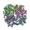
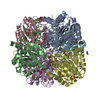
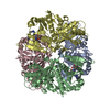
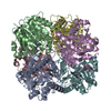
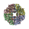
- PDB-7lox: The structure of Agmatinase from E. Coli at 3.2 A displaying guan... -
+ Open data

- Basic information
| Entry | Database: PDB / ID: 7lox | ||||||||||||
|---|---|---|---|---|---|---|---|---|---|---|---|---|---|
| Title | The structure of Agmatinase from E. Coli at 3.2 A displaying guanidine in the active site | ||||||||||||
 Components Components | Agmatinase | ||||||||||||
 Keywords Keywords | HYDROLASE | ||||||||||||
| Function / homology |  Function and homology information Function and homology informationagmatinase / agmatinase activity / : / spermidine biosynthetic process / manganese ion binding Similarity search - Function | ||||||||||||
| Biological species |  | ||||||||||||
| Method |  X-RAY DIFFRACTION / SYNCHROTRON / MOLECULAR REPLACEMENT / Resolution: 3.2 Å X-RAY DIFFRACTION / SYNCHROTRON / MOLECULAR REPLACEMENT / Resolution: 3.2 Å | ||||||||||||
 Authors Authors | Maturana, P. / Figueroa, M. / Gonzalez-Ordenes, F. / Villalobos, P. / Martinez-Oyanedel, J. / Uribe, E.A. / Castro-Fernandez, V. | ||||||||||||
| Funding support |  Chile, 3items Chile, 3items
| ||||||||||||
 Citation Citation | Journal: Int J Mol Sci / Year: 2021 Title: Crystal Structure of Escherichia coli Agmatinase: Catalytic Mechanism and Residues Relevant for Substrate Specificity. Authors: Maturana, P. / Orellana, M.S. / Herrera, S.M. / Martinez, I. / Figueroa, M. / Martinez-Oyanedel, J. / Castro-Fernandez, V. / Uribe, E. | ||||||||||||
| History |
|
- Structure visualization
| Structure viewer | Molecule: MolmilJmol/JSmol |
|---|
- Downloads & links
-Download
| PDBx/mmCIF format | 7lox.cif.gz | 189.1 KB | Display | PDBx/mmCIF format |
|---|---|---|---|---|
| PDB format | pdb7lox.ent.gz | 137.1 KB | Display | PDB format |
| PDBx/mmJSON format | 7lox.json.gz | Tree view | PDBx/mmJSON format | |
| Others |  Other downloads Other downloads |
-Validation report
| Arichive directory | https://data.pdbj.org/pub/pdb/validation_reports/lo/7loxftp://data.pdbj.org/pub/pdb/validation_reports/lo/7lox | HTTPS FTP |
|---|
-Related structure data
| Related structure data |  7lolC 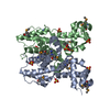 3npiS C: citing same article ( S: Starting model for refinement |
|---|---|
| Similar structure data |


-Links
 PDBj
PDBj- Assembly
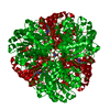
| Deposited unit | 
| |||||||||||||||||||||||||||||||||||||||||||||||||||||||||||||||||||||||||||||||||||||
|---|---|---|---|---|---|---|---|---|---|---|---|---|---|---|---|---|---|---|---|---|---|---|---|---|---|---|---|---|---|---|---|---|---|---|---|---|---|---|---|---|---|---|---|---|---|---|---|---|---|---|---|---|---|---|---|---|---|---|---|---|---|---|---|---|---|---|---|---|---|---|---|---|---|---|---|---|---|---|---|---|---|---|---|---|---|---|
| 1 | 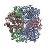
| |||||||||||||||||||||||||||||||||||||||||||||||||||||||||||||||||||||||||||||||||||||
| Unit cell |
| |||||||||||||||||||||||||||||||||||||||||||||||||||||||||||||||||||||||||||||||||||||
| Noncrystallographic symmetry (NCS) | NCS domain:
NCS domain segments: Ens-ID: 1
|
-Components
| #1: Protein | Mass: 33593.988 Da / Num. of mol.: 3 Source method: isolated from a genetically manipulated source Source: (gene. exp.) #2: Chemical | ChemComp-MN /   Mass: 54.938 Da / Num. of mol.: 6 / Source method: obtained synthetically / Formula: Mn / Feature type: SUBJECT OF INVESTIGATION Mass: 54.938 Da / Num. of mol.: 6 / Source method: obtained synthetically / Formula: Mn / Feature type: SUBJECT OF INVESTIGATION#3: Chemical | ChemComp-GAI / 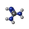  Mass: 59.070 Da / Num. of mol.: 4 / Source method: obtained synthetically / Formula: CH5N3 / Feature type: SUBJECT OF INVESTIGATION Mass: 59.070 Da / Num. of mol.: 4 / Source method: obtained synthetically / Formula: CH5N3 / Feature type: SUBJECT OF INVESTIGATIONHas ligand of interest | Y | |
|---|
-Experimental details
-Experiment
| Experiment | Method: X-RAY DIFFRACTION / Number of used crystals: 1 |
|---|
- Sample preparation
| Crystal | Density Matthews: 2.11 Å3/Da / Density % sol: 41.57 % |
|---|---|
| Crystal grow | Temperature: 291 K / Method: vapor diffusion, sitting drop / pH: 7.5 Details: Protein at 12 mg/mL in Buffer 25 mM Tris-HCl pH 8.0, 2 mM MnCl. Crystallization: 0.2 M Lithium sulfate monohydrate, 0.1 M HEPES pH 7.5, 25% w/v Polyethylene glycol 3350 |
-Data collection
| Diffraction | Mean temperature: 100 K / Serial crystal experiment: N |
|---|---|
| Diffraction source | Source: SYNCHROTRON / Site: APS  / Beamline: 23-ID-B / Wavelength: 1.771 Å / Beamline: 23-ID-B / Wavelength: 1.771 Å |
| Detector | Type: DECTRIS EIGER X 16M / Detector: PIXEL / Date: Dec 10, 2019 |
| Radiation | Protocol: SINGLE WAVELENGTH / Monochromatic (M) / Laue (L): M / Scattering type: x-ray |
| Radiation wavelength | Wavelength: 1.771 Å / Relative weight: 1 |
| Reflection | Resolution: 3.2→64.51 Å / Num. obs: 14314 / % possible obs: 99.87 % / Redundancy: 39.4 % / Biso Wilson estimate: 84.61 Å2 / Rmerge(I) obs: 0.1561 / Rrim(I) all: 0.1582 / Net I/σ(I): 26.7 |
| Reflection shell | Resolution: 3.2→3.314 Å / Rmerge(I) obs: 0.1561 / Num. unique obs: 1418 / Rrim(I) all: 0.1582 / % possible all: 100 |
- Processing
| Software |
| ||||||||||||||||||||||||||||||||||||||||||||||||||||||||||||||||||||||
|---|---|---|---|---|---|---|---|---|---|---|---|---|---|---|---|---|---|---|---|---|---|---|---|---|---|---|---|---|---|---|---|---|---|---|---|---|---|---|---|---|---|---|---|---|---|---|---|---|---|---|---|---|---|---|---|---|---|---|---|---|---|---|---|---|---|---|---|---|---|---|---|
| Refinement | Method to determine structure: MOLECULAR REPLACEMENT Starting model: 3NPI Resolution: 3.2→64.51 Å / SU ML: 0.292 / Cross valid method: FREE R-VALUE / σ(F): 1.34 / Phase error: 21.8165 / Stereochemistry target values: CDL v1.2
| ||||||||||||||||||||||||||||||||||||||||||||||||||||||||||||||||||||||
| Solvent computation | Shrinkage radii: 0.9 Å / VDW probe radii: 1.11 Å / Solvent model: FLAT BULK SOLVENT MODEL | ||||||||||||||||||||||||||||||||||||||||||||||||||||||||||||||||||||||
| Displacement parameters | Biso mean: 81.5 Å2 | ||||||||||||||||||||||||||||||||||||||||||||||||||||||||||||||||||||||
| Refinement step | Cycle: LAST / Resolution: 3.2→64.51 Å
| ||||||||||||||||||||||||||||||||||||||||||||||||||||||||||||||||||||||
| Refine LS restraints |
| ||||||||||||||||||||||||||||||||||||||||||||||||||||||||||||||||||||||
| LS refinement shell |
|
