 Movie
Movie Controller


+ Open data

- Basic information
| Entry | Database: PDB / ID: 7lba | ||||||||||||
|---|---|---|---|---|---|---|---|---|---|---|---|---|---|
| Title | E. coli Agmatinase | ||||||||||||
 Components Components | Agmatinase | ||||||||||||
 Keywords Keywords | HYDROLASE / Agmatine Polyamine amidinohydrolase ureohydrolase metallohydrolase | ||||||||||||
| Function / homology |  Function and homology information Function and homology informationagmatinase / agmatinase activity / : / spermidine biosynthetic process / manganese ion binding Similarity search - Function | ||||||||||||
| Biological species |  | ||||||||||||
| Method |  X-RAY DIFFRACTION / SYNCHROTRON / MOLECULAR REPLACEMENT / Resolution: 2.2 Å X-RAY DIFFRACTION / SYNCHROTRON / MOLECULAR REPLACEMENT / Resolution: 2.2 Å | ||||||||||||
 Authors Authors | Chitrakar, I. / Ahmed, S.F. / Torelli, A.T. / French, J.B. | ||||||||||||
| Funding support |  United States, 3items United States, 3items
| ||||||||||||
 Citation Citation | Journal: Plos One / Year: 2021 Title: Structure of the E. coli agmatinase, SPEB. Authors: Chitrakar, I. / Ahmed, S.F. / Torelli, A.T. / French, J.B. | ||||||||||||
| History |
|
- Structure visualization
| Structure viewer | Molecule: MolmilJmol/JSmol |
|---|
- Downloads & links
-Download
| PDBx/mmCIF format | 7lba.cif.gz | 1022.6 KB | Display | PDBx/mmCIF format |
|---|---|---|---|---|
| PDB format | pdb7lba.ent.gz | 846 KB | Display | PDB format |
| PDBx/mmJSON format | 7lba.json.gz | Tree view | PDBx/mmJSON format | |
| Others |  Other downloads Other downloads |
-Validation report
| Arichive directory | https://data.pdbj.org/pub/pdb/validation_reports/lb/7lbaftp://data.pdbj.org/pub/pdb/validation_reports/lb/7lba | HTTPS FTP |
|---|
-Related structure data
| Related structure data | 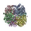 4dz4S S: Starting model for refinement |
|---|---|
| Similar structure data |


-Links
 PDBj
PDBj- Assembly
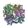
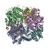
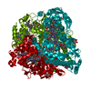
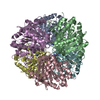
| Deposited unit | 
| ||||||||
|---|---|---|---|---|---|---|---|---|---|
| 1 | 
| ||||||||
| 2 | 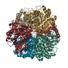
| ||||||||
| 3 | 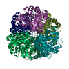
| ||||||||
| Unit cell |
|
-Components
| #1: Protein | Mass: 36276.859 Da / Num. of mol.: 18 Source method: isolated from a genetically manipulated source Source: (gene. exp.) #2: Chemical | ChemComp-MN /   Mass: 54.938 Da / Num. of mol.: 27 / Source method: obtained synthetically / Formula: Mn Mass: 54.938 Da / Num. of mol.: 27 / Source method: obtained synthetically / Formula: Mn#3: Water | ChemComp-HOH / |  Mass: 18.015 Da / Num. of mol.: 1249 / Source method: isolated from a natural source / Formula: H2O Mass: 18.015 Da / Num. of mol.: 1249 / Source method: isolated from a natural source / Formula: H2OHas ligand of interest | N | |
|---|
-Experimental details
-Experiment
| Experiment | Method: X-RAY DIFFRACTION / Number of used crystals: 1 |
|---|
- Sample preparation
| Crystal | Density Matthews: 2.98 Å3/Da / Density % sol: 58.8 % |
|---|---|
| Crystal grow | Temperature: 291 K / Method: vapor diffusion, hanging drop / pH: 8.5 / Details: 3.5 M Sodium Formate, 0.1 M Tris, pH 8.5 |
-Data collection
| Diffraction | Mean temperature: 100 K / Serial crystal experiment: N | ||||||||||||||||||||||||||||||
|---|---|---|---|---|---|---|---|---|---|---|---|---|---|---|---|---|---|---|---|---|---|---|---|---|---|---|---|---|---|---|---|
| Diffraction source | Source: SYNCHROTRON / Site: APS / Beamline: 24-ID-C / Wavelength: 0.97918 Å | ||||||||||||||||||||||||||||||
| Detector | Type: DECTRIS PILATUS3 6M / Detector: PIXEL / Date: Feb 16, 2018 | ||||||||||||||||||||||||||||||
| Radiation | Protocol: SINGLE WAVELENGTH / Monochromatic (M) / Laue (L): M / Scattering type: x-ray | ||||||||||||||||||||||||||||||
| Radiation wavelength | Wavelength: 0.97918 Å / Relative weight: 1 | ||||||||||||||||||||||||||||||
| Reflection twin |
| ||||||||||||||||||||||||||||||
| Reflection | Resolution: 2.2→82.02 Å / Num. obs: 247407 / % possible obs: 99.9 % / Redundancy: 5.8 % / CC1/2: 0.996 / Rmerge(I) obs: 0.123 / Rpim(I) all: 0.056 / Rrim(I) all: 0.136 / Net I/σ(I): 11.2 / Num. measured all: 1435676 / Scaling rejects: 254 | ||||||||||||||||||||||||||||||
| Reflection shell | Diffraction-ID: 1
|
- Processing
| Software |
| ||||||||||||||||||||||||||||||||||||||||||||||||||||||||||||
|---|---|---|---|---|---|---|---|---|---|---|---|---|---|---|---|---|---|---|---|---|---|---|---|---|---|---|---|---|---|---|---|---|---|---|---|---|---|---|---|---|---|---|---|---|---|---|---|---|---|---|---|---|---|---|---|---|---|---|---|---|---|
| Refinement | Method to determine structure: MOLECULAR REPLACEMENT Starting model: 4DZ4 Resolution: 2.2→82.02 Å / Cor.coef. Fo:Fc: 0.939 / Cor.coef. Fo:Fc free: 0.916 / SU B: 5.139 / SU ML: 0.136 / Cross valid method: THROUGHOUT / σ(F): 0 / ESU R: 0.072 / ESU R Free: 0.044 / Stereochemistry target values: MAXIMUM LIKELIHOOD Details: HYDROGENS HAVE BEEN ADDED IN THE RIDING POSITIONS U VALUES : REFINED INDIVIDUALLY
| ||||||||||||||||||||||||||||||||||||||||||||||||||||||||||||
| Solvent computation | Ion probe radii: 0.8 Å / Shrinkage radii: 0.8 Å / VDW probe radii: 1.2 Å / Solvent model: MASK | ||||||||||||||||||||||||||||||||||||||||||||||||||||||||||||
| Displacement parameters | Biso max: 108.77 Å2 / Biso mean: 27.061 Å2 / Biso min: 0.5 Å2
| ||||||||||||||||||||||||||||||||||||||||||||||||||||||||||||
| Refinement step | Cycle: final / Resolution: 2.2→82.02 Å
| ||||||||||||||||||||||||||||||||||||||||||||||||||||||||||||
| Refine LS restraints |
| ||||||||||||||||||||||||||||||||||||||||||||||||||||||||||||
| LS refinement shell | Resolution: 2.2→2.257 Å / Rfactor Rfree error: 0 / Total num. of bins used: 20
|
