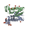
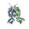
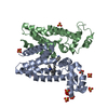
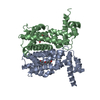
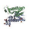
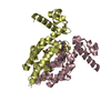
ANALYTICAL SIZE EXCLUSION CHROMATOGRAPHY WITH STATIC LIGHT SCATTERING SUPPORTS THE ASSIGNMENT OF A DIMER AS A SIGNIFICANT OLIGOMERIZATION STATE IN SOLUTION.
-
Components
#1: Protein
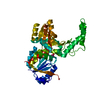
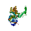
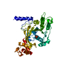
TetRfamilyregulatoryprotein
Mass: 28217.109 Da / Num. of mol.: 2 Source method: isolated from a genetically manipulated source Source: (gene. exp.) Corynebacterium diphtheriae (bacteria) / Gene: DIP1788 / Plasmid: SpeedET / Production host: Escherichia Coli (E. coli) / Strain (production host): HK100 / References: UniProt: Q6NFU8
Mass: 96.063 Da / Num. of mol.: 8 / Source method: obtained synthetically / Formula: SO4
Has protein modification
Y
Sequence details
THIS CONSTRUCT WAS EXPRESSED WITH A PURIFICATION TAG MGSDKIHHHHHHENLYFQG. THE TAG WAS REMOVED WITH ...THIS CONSTRUCT WAS EXPRESSED WITH A PURIFICATION TAG MGSDKIHHHHHHENLYFQG. THE TAG WAS REMOVED WITH TEV PROTEASE LEAVING ONLY A GLYCINE (0) FOLLOWED BY THE TARGET SEQUENCE.
-
Experimental details
-
Experiment
Experiment
Method: X-RAY DIFFRACTION / Number of used crystals: 1
-
Sample preparation
Crystal
Density Matthews: 2.64 Å3/Da / Density % sol: 53.34 %
Crystal grow
Temperature: 277 K / Method: vapor diffusion, sitting drop / pH: 6.86 Details: 1.7100M ammonium sulfate, 10.0000% Dioxane, 0.1M MES pH 6.86, VAPOR DIFFUSION,SITTING DROP,NANODROP, temperature 277K, VAPOR DIFFUSION, SITTING DROP
Type: MARMOSAIC 325 mm CCD / Detector: CCD / Date: Feb 11, 2010 / Details: Flat mirror (vertical focusing)
Radiation
Monochromator: Single crystal Si(111) bent monochromator (horizontal focusing) Protocol: MAD / Monochromatic (M) / Laue (L): M / Scattering type: x-ray
Radiation wavelength
ID
Wavelength (Å)
Relative weight
1
0.91837
1
2
0.97959
1
3
0.97925
1
Reflection
Resolution: 2.96→29.713 Å / Num. obs: 12584 / % possible obs: 99.7 % / Observed criterion σ(I): -3 / Biso Wilson estimate: 99.731 Å2 / Rmerge F obs: 0.115 / Rmerge(I) obs: 0.056 / Rrim(I) all: 0.066 / Net I/σ(I): 17.06 / Num. measured all: 93470
Reflection shell
Diffraction-ID: 1
Resolution (Å)
Highest resolution (Å)
Rmerge F obs
Rmerge(I) obs
Mean I/σ(I) obs
Num. measured obs
Num. possible
Num. unique obs
Rrim(I) all
% possible all
2.97-3.08
0.947
0.743
1.8
9695
2501
2496
0.863
99.8
3.08-3.2
0.623
0.485
2.8
9115
2355
2351
0.563
99.8
3.2-3.34
0.354
0.305
4.4
8872
2277
2277
0.354
100
3.34-3.52
0.249
0.201
6.6
9641
2481
2480
0.234
100
3.52-3.74
0.14
0.121
10.6
9257
2379
2374
0.14
99.8
3.74-4.02
0.09
0.077
15.8
9179
2357
2355
0.089
99.9
4.02-4.43
0.051
0.049
23.6
9568
2456
2451
0.056
99.8
4.43-5.06
0.036
0.04
29.4
9309
2393
2378
0.046
99.4
5.06-6.34
0.037
0.039
29.2
9275
2381
2377
0.046
99.8
6.34
0.018
0.024
45.8
9559
2490
2455
0.028
98.6
-
Phasing
Phasing
Method: MAD
-
Processing
Software
Name
Version
Classification
NB
SHELX
phasing
BUSTER-TNT
BUSTER2.8.0
refinement
XSCALE
dataprocessing
PDB_EXTRACT
3.1
dataextraction
XDS
datareduction
XSCALE
datascaling
autoSHARP
phasing
BUSTER
2.8.0
refinement
Refinement
Method to determine structure: MAD / Resolution: 2.96→29.713 Å / Cor.coef. Fo:Fc: 0.9434 / Cor.coef. Fo:Fc free: 0.9233 / Occupancy max: 1 / Occupancy min: 0.5 / Cross valid method: THROUGHOUT / σ(F): 0 Details: 1.A MET-INHIBITION PROTOCOL WAS USED FOR SELENOMETHIONINE INCORPORATION DURING PROTEIN EXPRESSION. THE OCCUPANCY OF THE SE ATOMS IN THE MSE RESIDUES WAS REDUCED TO 0.75 FOR THE REDUCED ...Details: 1.A MET-INHIBITION PROTOCOL WAS USED FOR SELENOMETHIONINE INCORPORATION DURING PROTEIN EXPRESSION. THE OCCUPANCY OF THE SE ATOMS IN THE MSE RESIDUES WAS REDUCED TO 0.75 FOR THE REDUCED SCATTERING POWER DUE TO PARTIAL S-MET INCORPORATION. 2. SULFATES FROM THE CRYSTALLIZATION WERE MODELED INTO THE STRUCTURE. 3.ATOM RECORD CONTAINS SUM OF TLS AND RESIDUAL B FACTORS. ANISOU RECORD CONTAINS SUM OF TLS AND RESIDUAL U FACTORS.
In the structure databanks used in Yorodumi, some data are registered as the other names, "COVID-19 virus" and "2019-nCoV". Here are the details of the virus and the list of structure data.
Jan 31, 2019. EMDB accession codes are about to change! (news from PDBe EMDB page)
EMDB accession codes are about to change! (news from PDBe EMDB page)
The allocation of 4 digits for EMDB accession codes will soon come to an end. Whilst these codes will remain in use, new EMDB accession codes will include an additional digit and will expand incrementally as the available range of codes is exhausted. The current 4-digit format prefixed with “EMD-” (i.e. EMD-XXXX) will advance to a 5-digit format (i.e. EMD-XXXXX), and so on. It is currently estimated that the 4-digit codes will be depleted around Spring 2019, at which point the 5-digit format will come into force.
The EM Navigator/Yorodumi systems omit the EMD- prefix.
Related info.:Q: What is EMD? / ID/Accession-code notation in Yorodumi/EM Navigator
Yorodumi is a browser for structure data from EMDB, PDB, SASBDB, etc.
This page is also the successor to EM Navigator detail page, and also detail information page/front-end page for Omokage search.
The word "yorodu" (or yorozu) is an old Japanese word meaning "ten thousand". "mi" (miru) is to see.
Related info.:EMDB / PDB / SASBDB / Comparison of 3 databanks / Yorodumi Search / Aug 31, 2016. New EM Navigator & Yorodumi / Yorodumi Papers / Jmol/JSmol / Function and homology information / Changes in new EM Navigator and Yorodumi
 Movie
Movie Controller

 Yorodumi
 Open data
 Basic information
 Components
 Keywords
 Function and homology information
 Corynebacterium diphtheriae (bacteria)
 X-RAY DIFFRACTION /
  Authors
 Citation
 Structure visualization
 Downloads & links
 Other downloads
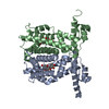
 PDBj
PDBj Assembly
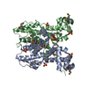

 Mass: 96.063 Da / Num. of mol.: 8 / Source method: obtained synthetically / Formula: SO4
 Sample preparation
 / Beamline: BL11-1 / Wavelength: 0.91837,0.97959,0.97925
 Processing