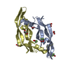
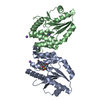
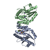
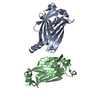
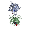
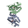
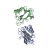
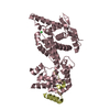
Entry Database : PDB / ID : 7ll8Title D-Protein RFX-V1 Bound to the VEGFR1 Domain 2 Site on VEGF-A Isoform L-VEGF189 of Vascular endothelial growth factor A RFX-V1 Keywords / / Function / homology Function Domain/homology Component
/ / / / / / / / / / / / / / / / / / / / / / / / / / / / / / / / / / / / / / / / / / / / / / / / / / / / / / / / / / / / / / / / / / / / / / / / / / / / / / / / / / / / / / / / / / / / / / / / / / / / / / / / / / / / / / / / / / / / / / Biological species Homo sapiens (human)synthetic construct (others) Method / / / Resolution : 2.31 Å Authors Marinec, P.S. / Landgraf, K.E. / Uppalapati, M. / Chen, G. / Xie, D. / Jiang, Q. / Zhao, Y. / Petriello, A. / Deshayes, K. / Kent, S.B.H. ...Marinec, P.S. / Landgraf, K.E. / Uppalapati, M. / Chen, G. / Xie, D. / Jiang, Q. / Zhao, Y. / Petriello, A. / Deshayes, K. / Kent, S.B.H. / Ault-Riche, D. / Sidhu, S.S. Journal : Acs Chem.Biol. / Year : 2021Title : A Non-immunogenic Bivalent d-Protein Potently Inhibits Retinal Vascularization and Tumor Growth.Authors : Marinec, P.S. / Landgraf, K.E. / Uppalapati, M. / Chen, G. / Xie, D. / Jiang, Q. / Zhao, Y. / Petriello, A. / Deshayes, K. / Kent, S.B.H. / Ault-Riche, D. / Sidhu, S.S. History Deposition Feb 3, 2021 Deposition site / Processing site Revision 1.0 Feb 17, 2021 Provider / Type Revision 1.1 Mar 10, 2021 Group / Category / citation_authorItem _citation.country / _citation.journal_abbrev ... _citation.country / _citation.journal_abbrev / _citation.journal_id_CSD / _citation.journal_id_ISSN / _citation.pdbx_database_id_DOI / _citation.pdbx_database_id_PubMed / _citation.title / _citation.year / _citation_author.identifier_ORCID Revision 1.2 Mar 31, 2021 Group / Category / citation_authorItem _citation.journal_volume / _citation.page_first ... _citation.journal_volume / _citation.page_first / _citation.page_last / _citation_author.identifier_ORCID Revision 1.3 Oct 18, 2023 Group / Database references / Refinement descriptionCategory chem_comp_atom / chem_comp_bond ... chem_comp_atom / chem_comp_bond / database_2 / pdbx_initial_refinement_model Item / _database_2.pdbx_database_accessionRevision 1.4 Nov 15, 2023 Group / Category / chem_comp_bond / Item / _chem_comp_bond.atom_id_2Revision 1.5 Nov 6, 2024 Group / Category / pdbx_modification_feature / Item
Show all Show less
 Movie
Movie Controller


 Open data
 Basic information
 Components
 Keywords
 Function and homology information
 Homo sapiens (human)
 X-RAY DIFFRACTION /
  Authors
 Citation
 Structure visualization
 Downloads & links
 Other downloads


 PDBj
PDBj


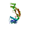
 Assembly
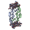

 Mass: 18.015 Da / Num. of mol.: 118 / Source method: isolated from a natural source / Formula: H2O
 Sample preparation
 / Beamline: BL19U1 / Wavelength: 0.9785 Å
 Processing