 Movie
Movie Controller

[English] 日本語
 Yorodumi
- PDB-7li0: Crystal structure of apo Moraxella catarrhalis ferric binding pro... -
+ Open data

- Basic information
| Entry | Database: PDB / ID: 7li0 | ||||||
|---|---|---|---|---|---|---|---|
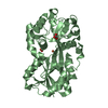
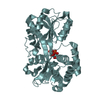
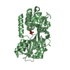
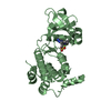
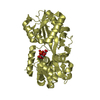
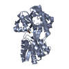
| Title | Crystal structure of apo Moraxella catarrhalis ferric binding protein A in an open conformation | ||||||
 Components Components | Fe(3+) ABC transporter substrate-binding protein | ||||||
 Keywords Keywords | METAL TRANSPORT / ferric binding protein A / FbpA | ||||||
| Function / homology | Ferric binding protein / Bacterial extracellular solute-binding protein / Bacterial extracellular solute-binding protein / outer membrane-bounded periplasmic space / metal ion binding / CITRIC ACID / CARBONATE ION / DI(HYDROXYETHYL)ETHER / Fe(3+) ABC transporter substrate-binding protein Function and homology information Function and homology information | ||||||
| Biological species |  Moraxella catarrhalis (bacteria) Moraxella catarrhalis (bacteria) | ||||||
| Method |  X-RAY DIFFRACTION / SYNCHROTRON / MOLECULAR REPLACEMENT / Resolution: 1.85 Å X-RAY DIFFRACTION / SYNCHROTRON / MOLECULAR REPLACEMENT / Resolution: 1.85 Å | ||||||
 Authors Authors | Chan, C. / Ng, D. / Fraser, M.E. / Schryvers, A.B. | ||||||
| Funding support |  Canada, 1items Canada, 1items
| ||||||
 Citation Citation | Journal: Biometals / Year: 2022 Title: Structural and functional insights into iron acquisition from lactoferrin and transferrin in Gram-negative bacterial pathogens. Authors: Chan, C. / Ng, D. / Fraser, M.E. / Schryvers, A.B. | ||||||
| History |
|
- Structure visualization
| Structure viewer | Molecule: MolmilJmol/JSmol |
|---|
- Downloads & links
-Download
| PDBx/mmCIF format | 7li0.cif.gz | 261.7 KB | Display | PDBx/mmCIF format |
|---|---|---|---|---|
| PDB format | pdb7li0.ent.gz | 206.7 KB | Display | PDB format |
| PDBx/mmJSON format | 7li0.json.gz | Tree view | PDBx/mmJSON format | |
| Others |  Other downloads Other downloads |
-Validation report
| Arichive directory | https://data.pdbj.org/pub/pdb/validation_reports/li/7li0ftp://data.pdbj.org/pub/pdb/validation_reports/li/7li0 | HTTPS FTP |
|---|
-Related structure data
| Related structure data | 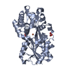 7li1C 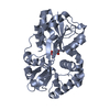 6g7nS S: Starting model for refinement C: citing same article ( |
|---|---|
| Similar structure data |


-Links
 PDBj
PDBj


- Assembly
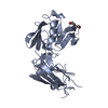
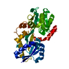
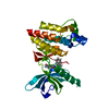
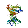
| Deposited unit | 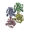
| ||||||||||||
|---|---|---|---|---|---|---|---|---|---|---|---|---|---|
| 1 | 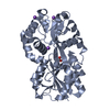
| ||||||||||||
| 2 | 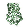
| ||||||||||||
| 3 | 
| ||||||||||||
| 4 | 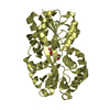
| ||||||||||||
| Unit cell |
|
-Components
-Protein , 1 types, 4 molecules ABCD
| #1: Protein | Mass: 34304.738 Da / Num. of mol.: 4 Source method: isolated from a genetically manipulated source Source: (gene. exp.) Moraxella catarrhalis (bacteria) / Gene: fbpA / Production host: |
|---|
-Non-polymers , 6 types, 734 molecules 


| #2: Chemical |  Mass: 60.009 Da / Num. of mol.: 3 / Source method: obtained synthetically / Formula: CO3 Mass: 60.009 Da / Num. of mol.: 3 / Source method: obtained synthetically / Formula: CO3#3: Chemical | ChemComp-NA /  Mass: 22.990 Da / Num. of mol.: 4 / Source method: obtained synthetically / Formula: Na Mass: 22.990 Da / Num. of mol.: 4 / Source method: obtained synthetically / Formula: Na#4: Chemical |  Mass: 92.094 Da / Num. of mol.: 2 / Source method: obtained synthetically / Formula: C3H8O3 Mass: 92.094 Da / Num. of mol.: 2 / Source method: obtained synthetically / Formula: C3H8O3#5: Chemical | ChemComp-PEG / |  Mass: 106.120 Da / Num. of mol.: 1 / Source method: obtained synthetically / Formula: C4H10O3 Mass: 106.120 Da / Num. of mol.: 1 / Source method: obtained synthetically / Formula: C4H10O3#6: Chemical | ChemComp-CIT / |  Mass: 192.124 Da / Num. of mol.: 1 / Source method: obtained synthetically / Formula: C6H8O7 Mass: 192.124 Da / Num. of mol.: 1 / Source method: obtained synthetically / Formula: C6H8O7#7: Water | ChemComp-HOH / | Mass: 18.015 Da / Num. of mol.: 723 / Source method: isolated from a natural source / Formula: H2O |
|---|
-Details
| Has ligand of interest | N |
|---|---|
| Has protein modification | Y |
-Experimental details
-Experiment
| Experiment | Method: X-RAY DIFFRACTION / Number of used crystals: 1 |
|---|
- Sample preparation
| Crystal | Density Matthews: 2.17 Å3/Da / Density % sol: 43.28 % |
|---|---|
| Crystal grow | Temperature: 298 K / Method: vapor diffusion, hanging drop / pH: 6.5 Details: 0.1 M ammonium citrate tribasic pH 6.5, 30% v/v polyethylene glycol 3350 |
-Data collection
| Diffraction | Mean temperature: 100 K / Serial crystal experiment: N |
|---|---|
| Diffraction source | Source: SYNCHROTRON / Site: NSLS-II  / Beamline: 17-ID-1 / Wavelength: 0.92009 Å / Beamline: 17-ID-1 / Wavelength: 0.92009 Å |
| Detector | Type: DECTRIS EIGER X 9M / Detector: PIXEL / Date: Nov 7, 2018 |
| Radiation | Protocol: SINGLE WAVELENGTH / Monochromatic (M) / Laue (L): M / Scattering type: x-ray |
| Radiation wavelength | Wavelength: 0.92009 Å / Relative weight: 1 |
| Reflection | Resolution: 1.84→51.61 Å / Num. obs: 96783 / % possible obs: 96.8 % / Redundancy: 1.8 % / CC1/2: 0.992 / Rmerge(I) obs: 0.058 / Net I/σ(I): 6.6 |
| Reflection shell | Resolution: 1.84→1.87 Å / Rmerge(I) obs: 0.416 / Num. unique obs: 4682 / CC1/2: 0.795 |
- Processing
| Software |
| ||||||||||||||||||||||||
|---|---|---|---|---|---|---|---|---|---|---|---|---|---|---|---|---|---|---|---|---|---|---|---|---|---|
| Refinement | Method to determine structure: MOLECULAR REPLACEMENT Starting model: 6G7N Resolution: 1.85→51.61 Å / Cross valid method: FREE R-VALUE Stereochemistry target values: GeoStd + Monomer Library + CDL v1.2
| ||||||||||||||||||||||||
| Displacement parameters | Biso mean: 34.67 Å2 | ||||||||||||||||||||||||
| Refinement step | Cycle: LAST / Resolution: 1.85→51.61 Å
| ||||||||||||||||||||||||
| Refine LS restraints |
|
