 Movie
Movie Controller

[English] 日本語
 Yorodumi
- PDB-7lfr: Crystal structure of the epidermal growth factor receptor extrace... -
+ Open data

- Basic information
| Entry | Database: PDB / ID: 7lfr | ||||||
|---|---|---|---|---|---|---|---|
| Title | Crystal structure of the epidermal growth factor receptor extracellular region with R84K mutation in complex with epiregulin crystallized with spermine | ||||||
 Components Components |
| ||||||
 Keywords Keywords | SIGNALING PROTEIN / receptor / epiregulin / glioblastoma / cancer / mutation / extracellular / asymmetric / dimer / ErbB1 / EGFR | ||||||
| Function / homology |  Function and homology information Function and homology informationprimary follicle stage / ovarian cumulus expansion / luteinizing hormone signaling pathway / negative regulation of smooth muscle cell differentiation / ERBB4-ERBB4 signaling pathway / ovulation / female meiotic nuclear division / keratinocyte proliferation / ERBB2-ERBB4 signaling pathway / transmembrane receptor protein tyrosine kinase activator activity ...primary follicle stage / ovarian cumulus expansion / luteinizing hormone signaling pathway / negative regulation of smooth muscle cell differentiation / ERBB4-ERBB4 signaling pathway / ovulation / female meiotic nuclear division / keratinocyte proliferation / ERBB2-ERBB4 signaling pathway / transmembrane receptor protein tyrosine kinase activator activity / oocyte maturation / PI3K events in ERBB4 signaling / positive regulation of innate immune response / multivesicular body, internal vesicle lumen / negative regulation of cardiocyte differentiation / Shc-EGFR complex / mRNA transcription / positive regulation of protein kinase C signaling / Inhibition of Signaling by Overexpressed EGFR / EGFR interacts with phospholipase C-gamma / epidermal growth factor receptor activity / epidermal growth factor binding / response to UV-A / epidermal growth factor receptor binding / regulation of peptidyl-tyrosine phosphorylation / PLCG1 events in ERBB2 signaling / ERBB2-EGFR signaling pathway / ERBB2 Activates PTK6 Signaling / PTK6 promotes HIF1A stabilization / Signaling by EGFR / intracellular vesicle / negative regulation of epidermal growth factor receptor signaling pathway / ERBB2 Regulates Cell Motility / positive regulation of protein kinase activity / Developmental Lineage of Mammary Gland Myoepithelial Cells / positive regulation of cell division / keratinocyte differentiation / protein insertion into membrane / Respiratory syncytial virus (RSV) attachment and entry / Signaling by ERBB4 / PI3K events in ERBB2 signaling / positive regulation of phosphorylation / positive regulation of peptidyl-serine phosphorylation / Estrogen-dependent nuclear events downstream of ESR-membrane signaling / MAP kinase kinase kinase activity / GAB1 signalosome / anatomical structure morphogenesis / protein tyrosine kinase activator activity / SHC1 events in ERBB4 signaling / Nuclear signaling by ERBB4 / ossification / positive regulation of G1/S transition of mitotic cell cycle / Signaling by ERBB2 / TFAP2 (AP-2) family regulates transcription of growth factors and their receptors / animal organ morphogenesis / positive regulation of mitotic nuclear division / transmembrane receptor protein tyrosine kinase activity / positive regulation of epithelial cell proliferation / GRB2 events in EGFR signaling / SHC1 events in EGFR signaling / positive regulation of smooth muscle cell proliferation / EGFR Transactivation by Gastrin / GRB2 events in ERBB2 signaling / positive regulation of fibroblast proliferation / SHC1 events in ERBB2 signaling / cellular response to epidermal growth factor stimulus / positive regulation of DNA repair / positive regulation of DNA replication / basal plasma membrane / cellular response to amino acid stimulus / positive regulation of cytokine production / wound healing / phosphatidylinositol 3-kinase/protein kinase B signal transduction / Signal transduction by L1 / cellular response to estradiol stimulus / positive regulation of protein localization to plasma membrane / growth factor activity / negative regulation of protein catabolic process / NOTCH3 Activation and Transmission of Signal to the Nucleus / clathrin-coated endocytic vesicle membrane / cell-cell adhesion / Signaling by ERBB2 TMD/JMD mutants / EGFR downregulation / Constitutive Signaling by EGFRvIII / receptor protein-tyrosine kinase / Signaling by ERBB2 ECD mutants / Signaling by ERBB2 KD Mutants / negative regulation of epithelial cell proliferation / positive regulation of interleukin-6 production / positive regulation of miRNA transcription / cell junction / positive regulation of protein phosphorylation / response to peptide hormone / epidermal growth factor receptor signaling pathway / kinase binding / ruffle membrane / cytokine-mediated signaling pathway / Downregulation of ERBB2 signaling / Constitutive Signaling by Aberrant PI3K in Cancer / HCMV Early Events Similarity search - Function | ||||||
| Biological species |  Homo sapiens (human) Homo sapiens (human) | ||||||
| Method |  X-RAY DIFFRACTION / SYNCHROTRON / MOLECULAR REPLACEMENT / Resolution: 3.2 Å X-RAY DIFFRACTION / SYNCHROTRON / MOLECULAR REPLACEMENT / Resolution: 3.2 Å | ||||||
 Authors Authors | Hu, C. / Leche II, C.A. / Stayrook, S.E. / Ferguson, K.M. / Lemmon, M.A. | ||||||
| Funding support |  United States, 1items United States, 1items
| ||||||
 Citation Citation | Journal: Nature / Year: 2022 Title: Glioblastoma mutations alter EGFR dimer structure to prevent ligand bias. Authors: Hu, C. / Leche 2nd, C.A. / Kiyatkin, A. / Yu, Z. / Stayrook, S.E. / Ferguson, K.M. / Lemmon, M.A. | ||||||
| History |
|
- Structure visualization
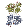
| Structure viewer | Molecule: MolmilJmol/JSmol |
|---|
- Downloads & links
-Download
| PDBx/mmCIF format | 7lfr.cif.gz | 223.2 KB | Display | PDBx/mmCIF format |
|---|---|---|---|---|
| PDB format | pdb7lfr.ent.gz | 176.1 KB | Display | PDB format |
| PDBx/mmJSON format | 7lfr.json.gz | Tree view | PDBx/mmJSON format | |
| Others |  Other downloads Other downloads |
-Validation report
| Arichive directory | https://data.pdbj.org/pub/pdb/validation_reports/lf/7lfrftp://data.pdbj.org/pub/pdb/validation_reports/lf/7lfr | HTTPS FTP |
|---|
-Related structure data
| Related structure data | 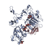 7lenC 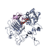 7lfsC 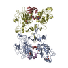 5wb7S C: citing same article ( S: Starting model for refinement |
|---|---|
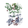
| Similar structure data |
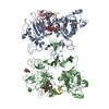
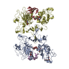
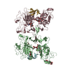
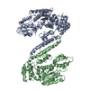

-Links
 PDBj
PDBj







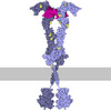

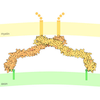

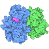
- Assembly
| Deposited unit | 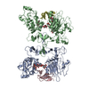
| ||||||||
|---|---|---|---|---|---|---|---|---|---|
| 1 |
| ||||||||
| 2 | 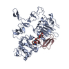
| ||||||||
| 3 | 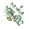
| ||||||||
| Unit cell |
|
-Components
-Protein / Protein/peptide , 2 types, 4 molecules ABCD
| #1: Protein | Mass: 55803.461 Da / Num. of mol.: 2 / Mutation: R84K Source method: isolated from a genetically manipulated source Source: (gene. exp.) Homo sapiens (human) / Gene: EGFR, ERBB, ERBB1, HER1 / Production host:   Spodoptera frugiperda (fall armyworm) Spodoptera frugiperda (fall armyworm)References: UniProt: P00533, receptor protein-tyrosine kinase #2: Protein/peptide | Mass: 5480.307 Da / Num. of mol.: 2 Source method: isolated from a genetically manipulated source Source: (gene. exp.) Homo sapiens (human) / Gene: EREG / Production host:  Drosophila melanogaster (fruit fly) / References: UniProt: O14944 Drosophila melanogaster (fruit fly) / References: UniProt: O14944 |
|---|
-Sugars , 4 types, 9 molecules 
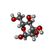
| #3: Polysaccharide | beta-D-mannopyranose-(1-4)-2-acetamido-2-deoxy-beta-D-glucopyranose | ||||
|---|---|---|---|---|---|
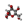
| #4: Sugar | ChemComp-NAG /  Type: D-saccharide, beta linking / Mass: 221.208 Da / Num. of mol.: 5 / Source method: obtained synthetically / Formula: C8H15NO6 Type: D-saccharide, beta linking / Mass: 221.208 Da / Num. of mol.: 5 / Source method: obtained synthetically / Formula: C8H15NO6#5: Sugar | 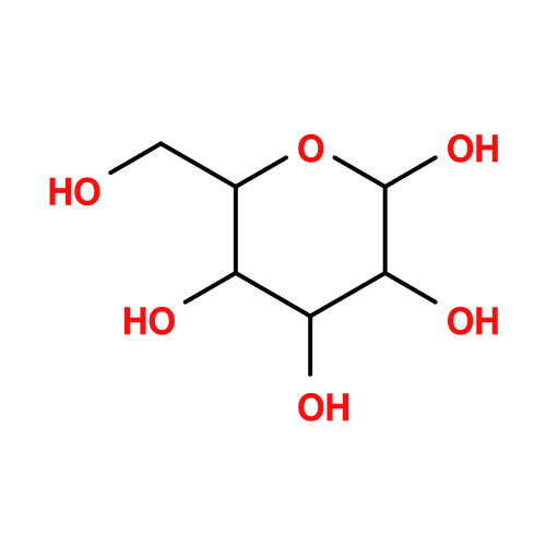 Type: D-saccharide, alpha linking / Mass: 180.156 Da / Num. of mol.: 2 / Source method: obtained synthetically / Formula: C6H12O6 Type: D-saccharide, alpha linking / Mass: 180.156 Da / Num. of mol.: 2 / Source method: obtained synthetically / Formula: C6H12O6#6: Sugar | ChemComp-BMA / | 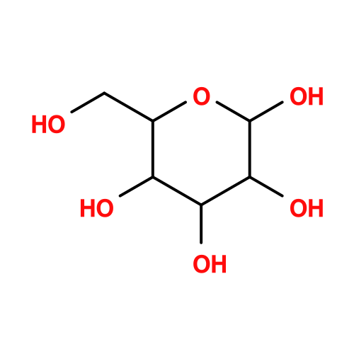 Type: D-saccharide, beta linking / Mass: 180.156 Da / Num. of mol.: 1 / Source method: obtained synthetically / Formula: C6H12O6 Type: D-saccharide, beta linking / Mass: 180.156 Da / Num. of mol.: 1 / Source method: obtained synthetically / Formula: C6H12O6 |
-Details
| Has ligand of interest | N |
|---|---|
| Has protein modification | Y |
-Experimental details
-Experiment
| Experiment | Method: X-RAY DIFFRACTION / Number of used crystals: 1 |
|---|
- Sample preparation
| Crystal | Density Matthews: 2.64 Å3/Da / Density % sol: 53 % |
|---|---|
| Crystal grow | Temperature: 293.15 K / Method: vapor diffusion, hanging drop / pH: 7.5 Details: 8 mg/ml protein, 100 mM HEPES (pH 7.5), 12% PEG3350, 10 mM spermine tetrahydrochloride |
-Data collection
| Diffraction | Mean temperature: 100 K / Serial crystal experiment: N | |||||||||||||||||||||||||||||||||||||||||||||||||||||||||||||||||||||||||||||||||||||||||||||||||||||||||||||||||||||||||
|---|---|---|---|---|---|---|---|---|---|---|---|---|---|---|---|---|---|---|---|---|---|---|---|---|---|---|---|---|---|---|---|---|---|---|---|---|---|---|---|---|---|---|---|---|---|---|---|---|---|---|---|---|---|---|---|---|---|---|---|---|---|---|---|---|---|---|---|---|---|---|---|---|---|---|---|---|---|---|---|---|---|---|---|---|---|---|---|---|---|---|---|---|---|---|---|---|---|---|---|---|---|---|---|---|---|---|---|---|---|---|---|---|---|---|---|---|---|---|---|---|---|---|
| Diffraction source | Source: SYNCHROTRON / Site: APS / Beamline: 23-ID-B / Wavelength: 1.0332 Å | |||||||||||||||||||||||||||||||||||||||||||||||||||||||||||||||||||||||||||||||||||||||||||||||||||||||||||||||||||||||||
| Detector | Type: DECTRIS EIGER X 16M / Detector: PIXEL / Date: Feb 14, 2020 / Details: Undulator | |||||||||||||||||||||||||||||||||||||||||||||||||||||||||||||||||||||||||||||||||||||||||||||||||||||||||||||||||||||||||
| Radiation | Protocol: SINGLE WAVELENGTH / Monochromatic (M) / Laue (L): M / Scattering type: x-ray | |||||||||||||||||||||||||||||||||||||||||||||||||||||||||||||||||||||||||||||||||||||||||||||||||||||||||||||||||||||||||
| Radiation wavelength | Wavelength: 1.0332 Å / Relative weight: 1 | |||||||||||||||||||||||||||||||||||||||||||||||||||||||||||||||||||||||||||||||||||||||||||||||||||||||||||||||||||||||||
| Reflection | Resolution: 3.2→99.006 Å / Num. obs: 22984 / % possible obs: 99.8 % / Redundancy: 8.4 % / Rpim(I) all: 0.073 / Rrim(I) all: 0.215 / Rsym value: 0.202 / Net I/av σ(I): 3.1 / Net I/σ(I): 9 / Num. measured all: 193914 | |||||||||||||||||||||||||||||||||||||||||||||||||||||||||||||||||||||||||||||||||||||||||||||||||||||||||||||||||||||||||
| Reflection shell | Diffraction-ID: 1
|
- Processing
| Software |
| |||||||||||||||||||||||||||||||||||||||||||||||||||||||||||||||
|---|---|---|---|---|---|---|---|---|---|---|---|---|---|---|---|---|---|---|---|---|---|---|---|---|---|---|---|---|---|---|---|---|---|---|---|---|---|---|---|---|---|---|---|---|---|---|---|---|---|---|---|---|---|---|---|---|---|---|---|---|---|---|---|---|
| Refinement | Method to determine structure: MOLECULAR REPLACEMENT Starting model: 5WB7 Resolution: 3.2→43.6 Å / SU ML: 0.47 / Cross valid method: THROUGHOUT / σ(F): 1.34 / Phase error: 30.43 / Stereochemistry target values: ML
| |||||||||||||||||||||||||||||||||||||||||||||||||||||||||||||||
| Solvent computation | Shrinkage radii: 0.9 Å / VDW probe radii: 1.11 Å / Solvent model: FLAT BULK SOLVENT MODEL | |||||||||||||||||||||||||||||||||||||||||||||||||||||||||||||||
| Displacement parameters | Biso max: 170.45 Å2 / Biso mean: 92.041 Å2 / Biso min: 40.83 Å2 | |||||||||||||||||||||||||||||||||||||||||||||||||||||||||||||||
| Refinement step | Cycle: final / Resolution: 3.2→43.6 Å
| |||||||||||||||||||||||||||||||||||||||||||||||||||||||||||||||
| LS refinement shell | Refine-ID: X-RAY DIFFRACTION / Rfactor Rfree error: 0 / Total num. of bins used: 8
|
