 Movie
Movie Controller


+ Open data

- Basic information
| Entry | Database: PDB / ID: 7lfd | ||||||
|---|---|---|---|---|---|---|---|
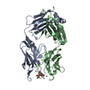
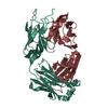
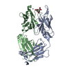
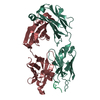
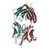
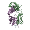
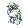
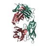
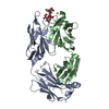
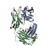
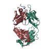
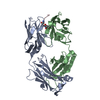
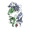
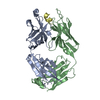
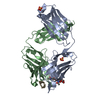
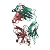
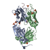
| Title | Fab 7D6 bound to ApoL1 BH3 like peptide | ||||||
 Components Components |
| ||||||
 Keywords Keywords | IMMUNE SYSTEM / Complex | ||||||
| Function / homology |  Function and homology information Function and homology informationlipoprotein metabolic process / high-density lipoprotein particle / very-low-density lipoprotein particle / : / lipid transport / chloride channel activity / Scavenging of heme from plasma / cholesterol metabolic process / chloride transmembrane transport / Post-translational protein phosphorylation ...lipoprotein metabolic process / high-density lipoprotein particle / very-low-density lipoprotein particle / : / lipid transport / chloride channel activity / Scavenging of heme from plasma / cholesterol metabolic process / chloride transmembrane transport / Post-translational protein phosphorylation / Regulation of Insulin-like Growth Factor (IGF) transport and uptake by Insulin-like Growth Factor Binding Proteins (IGFBPs) / blood microparticle / endoplasmic reticulum lumen / innate immune response / lipid binding / : / extracellular region / membrane Similarity search - Function | ||||||
| Biological species |  Homo sapiens (human) Homo sapiens (human) | ||||||
| Method |  X-RAY DIFFRACTION / SYNCHROTRON / MOLECULAR REPLACEMENT / Resolution: 2.157 Å X-RAY DIFFRACTION / SYNCHROTRON / MOLECULAR REPLACEMENT / Resolution: 2.157 Å | ||||||
 Authors Authors | Ultsch, M. / Kirchhofer, D. | ||||||
 Citation Citation | Journal: Commun Biol / Year: 2021 Title: Structures of the ApoL1 and ApoL2 N-terminal domains reveal a non-classical four-helix bundle motif. Authors: Ultsch, M. / Holliday, M.J. / Gerhardy, S. / Moran, P. / Scales, S.J. / Gupta, N. / Oltrabella, F. / Chiu, C. / Fairbrother, W. / Eigenbrot, C. / Kirchhofer, D. | ||||||
| History |
|
- Structure visualization
| Structure viewer | Molecule: MolmilJmol/JSmol |
|---|
- Downloads & links
-Download
| PDBx/mmCIF format | 7lfd.cif.gz | 199.2 KB | Display | PDBx/mmCIF format |
|---|---|---|---|---|
| PDB format | pdb7lfd.ent.gz | 158 KB | Display | PDB format |
| PDBx/mmJSON format | 7lfd.json.gz | Tree view | PDBx/mmJSON format | |
| Others |  Other downloads Other downloads |
-Validation report
| Arichive directory | https://data.pdbj.org/pub/pdb/validation_reports/lf/7lfdftp://data.pdbj.org/pub/pdb/validation_reports/lf/7lfd | HTTPS FTP |
|---|
-Related structure data
| Related structure data |  7l6kC  7lf7C 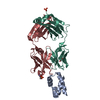 7lf8C  7lfaC  7lfbSC S: Starting model for refinement C: citing same article ( |
|---|---|
| Similar structure data |


-Links
 PDBj
PDBj


- Assembly
| Deposited unit | 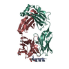
| ||||||||
|---|---|---|---|---|---|---|---|---|---|
| 1 |
| ||||||||
| Unit cell |
|
-Components
-Protein/peptide , 1 types, 1 molecules A
| #1: Protein/peptide | Mass: 1957.240 Da / Num. of mol.: 1 / Source method: obtained synthetically / Source: (synth.) Homo sapiens (human) / References: UniProt: O14791 |
|---|
-Antibody , 2 types, 2 molecules HL
| #2: Antibody | Mass: 24050.879 Da / Num. of mol.: 1 Source method: isolated from a genetically manipulated source Source: (gene. exp.) Homo sapiens (human) / Plasmid: AEP1 / Production host:  |
|---|---|
| #3: Antibody | Mass: 23325.795 Da / Num. of mol.: 1 Source method: isolated from a genetically manipulated source Source: (gene. exp.) Homo sapiens (human) / Plasmid: AEP1 / Production host: |
-Non-polymers , 5 types, 191 molecules 


| #4: Chemical | ChemComp-FLC /  Mass: 189.100 Da / Num. of mol.: 1 / Source method: obtained synthetically / Formula: C6H5O7 Mass: 189.100 Da / Num. of mol.: 1 / Source method: obtained synthetically / Formula: C6H5O7 | ||||
|---|---|---|---|---|---|
| #5: Chemical | ChemComp-EOH /  Mass: 46.068 Da / Num. of mol.: 1 / Source method: obtained synthetically / Formula: C2H6O Mass: 46.068 Da / Num. of mol.: 1 / Source method: obtained synthetically / Formula: C2H6O | ||||
| #6: Chemical |  Mass: 92.094 Da / Num. of mol.: 3 / Source method: obtained synthetically / Formula: C3H8O3 Mass: 92.094 Da / Num. of mol.: 3 / Source method: obtained synthetically / Formula: C3H8O3#7: Chemical | ChemComp-NH4 / |  Mass: 18.038 Da / Num. of mol.: 1 / Source method: obtained synthetically / Formula: H4N Mass: 18.038 Da / Num. of mol.: 1 / Source method: obtained synthetically / Formula: H4N#8: Water | ChemComp-HOH / | Mass: 18.015 Da / Num. of mol.: 185 / Source method: isolated from a natural source / Formula: H2O |
-Details
| Has ligand of interest | N |
|---|---|
| Has protein modification | Y |
-Experimental details
-Experiment
| Experiment | Method: X-RAY DIFFRACTION / Number of used crystals: 1 |
|---|
- Sample preparation
| Crystal | Description: rods |
|---|---|
| Crystal grow | Temperature: 291 K / Method: vapor diffusion, sitting drop / pH: 7 / Details: 1.8M Ammonium Citrate pH 7.0 / PH range: 6.5 - 7.5 |
-Data collection
| Diffraction | Mean temperature: 93 K / Serial crystal experiment: N |
|---|---|
| Diffraction source | Source: SYNCHROTRON / Site: ALS  / Beamline: 5.0.2 / Wavelength: 1 Å / Beamline: 5.0.2 / Wavelength: 1 Å |
| Detector | Type: DECTRIS PILATUS 300K / Detector: PIXEL / Date: Sep 20, 2018 |
| Radiation | Protocol: SINGLE WAVELENGTH / Monochromatic (M) / Laue (L): M / Scattering type: x-ray |
| Radiation wavelength | Wavelength: 1 Å / Relative weight: 1 |
| Reflection | Resolution: 2.157→70.674 Å / Num. obs: 40614 / % possible obs: 89.6 % / Redundancy: 9.4 % / CC1/2: 0.998 / Rmerge(I) obs: 0.149 / Rpim(I) all: 0.074 / Rrim(I) all: 0.167 / Net I/σ(I): 11.3 |
| Reflection shell | Resolution: 2.163→2.273 Å / Rmerge(I) obs: 1.638 / Mean I/σ(I) obs: 1.2 / Num. unique obs: 2033 / CC1/2: 0.371 / Rpim(I) all: 0.855 / Rrim(I) all: 1.855 |
- Processing
| Software |
| ||||||||||||||||||||||||||||||||||||||||||||||||||||||||||||||||||||||||||||||||||||||||||||||||||||||
|---|---|---|---|---|---|---|---|---|---|---|---|---|---|---|---|---|---|---|---|---|---|---|---|---|---|---|---|---|---|---|---|---|---|---|---|---|---|---|---|---|---|---|---|---|---|---|---|---|---|---|---|---|---|---|---|---|---|---|---|---|---|---|---|---|---|---|---|---|---|---|---|---|---|---|---|---|---|---|---|---|---|---|---|---|---|---|---|---|---|---|---|---|---|---|---|---|---|---|---|---|---|---|---|
| Refinement | Method to determine structure: MOLECULAR REPLACEMENT Starting model: 7LFB Resolution: 2.157→70.674 Å / SU ML: 0.28 / Cross valid method: THROUGHOUT / σ(F): 1.35 / Phase error: 23.7 / Stereochemistry target values: ML
| ||||||||||||||||||||||||||||||||||||||||||||||||||||||||||||||||||||||||||||||||||||||||||||||||||||||
| Solvent computation | Shrinkage radii: 0.9 Å / VDW probe radii: 1.11 Å / Solvent model: FLAT BULK SOLVENT MODEL | ||||||||||||||||||||||||||||||||||||||||||||||||||||||||||||||||||||||||||||||||||||||||||||||||||||||
| Displacement parameters | Biso max: 178.8 Å2 / Biso mean: 46.5584 Å2 / Biso min: 22.1 Å2 | ||||||||||||||||||||||||||||||||||||||||||||||||||||||||||||||||||||||||||||||||||||||||||||||||||||||
| Refinement step | Cycle: final / Resolution: 2.157→70.674 Å
| ||||||||||||||||||||||||||||||||||||||||||||||||||||||||||||||||||||||||||||||||||||||||||||||||||||||
| Refine LS restraints |
| ||||||||||||||||||||||||||||||||||||||||||||||||||||||||||||||||||||||||||||||||||||||||||||||||||||||
| LS refinement shell | Refine-ID: X-RAY DIFFRACTION / Rfactor Rfree error: 0
| ||||||||||||||||||||||||||||||||||||||||||||||||||||||||||||||||||||||||||||||||||||||||||||||||||||||
| Refinement TLS params. | Method: refined / Origin x: 115.9727 Å / Origin y: 82.7508 Å / Origin z: 6.8589 Å
| ||||||||||||||||||||||||||||||||||||||||||||||||||||||||||||||||||||||||||||||||||||||||||||||||||||||
| Refinement TLS group |
|
